• Laser & Optoelectronics Progress
  • Vol. 59, Issue 6, 0617025 (2022)
Na Fang1, Zanyi Wu2, Xingfu Wang3, Yuanxiang Lin2、**, and Jianxin Chen4、*
Author Affiliations
  • 1School of Medical Technology and Engineering, Fujian Medical University, Fuzhou , Fujian 350004, China
  • 2Department of Neurosurgery, the First Affiliated Hospital, Fujian Medical University, Fuzhou , Fujian 350004, China
  • 3Department of Pathology, the First Affiliated Hospital, Fujian Medical University, Fuzhou , Fujian 350004, China
  • 4Key Laboratory of OptoElectronic Science and Technology for Medicine, Ministry of Education, Fujian Normal University, Fuzhou , Fujian 350007, China
  • show less
    DOI: 10.3788/LOP202259.0617025 Cite this Article Set citation alerts
    Na Fang, Zanyi Wu, Xingfu Wang, Yuanxiang Lin, Jianxin Chen. Multiphoton Technique for Visualization of Angiomatous Meningiomas[J]. Laser & Optoelectronics Progress, 2022, 59(6): 0617025 Copy Citation Text show less
    Multi-photon spectral analysis of angiomatous meningiomas. (a) Normalized multi-photon emission spectrum; (b) multi-peak fitting
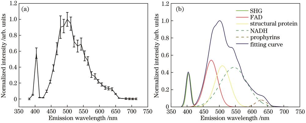
    Fig. 1. Multi-photon spectral analysis of angiomatous meningiomas. (a) Normalized multi-photon emission spectrum; (b) multi-peak fitting
    MPM images and the corresponding H&E stained image of normal dura mater and arachnoid mater. (a) SHG image; (b) TPEF image; (c) TPEF/SHG overlaid image; (d) corresponding H&E stained image
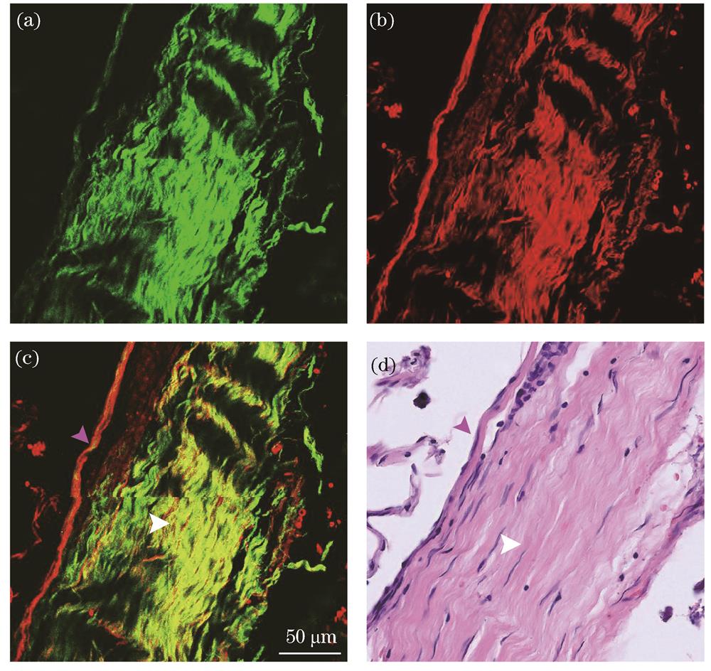
    Fig. 2. MPM images and the corresponding H&E stained image of normal dura mater and arachnoid mater. (a) SHG image; (b) TPEF image; (c) TPEF/SHG overlaid image; (d) corresponding H&E stained image
    MPM images and the corresponding H&E stained image of angiomatous meningioma. (a) TPEF image; (b) SHG image; (c) TPEF/SHG overlaid images; (d) corresponding H&E stained image
    Fig. 3. MPM images and the corresponding H&E stained image of angiomatous meningioma. (a) TPEF image; (b) SHG image; (c) TPEF/SHG overlaid images; (d) corresponding H&E stained image
    The enlarged images of the solid line box in Fig.3(c). (a) TPEF image; (b) SHG image; (c) TPEF/SHG overlaid image; (d) corresponding H&E stained image
    Fig. 4. The enlarged images of the solid line box in Fig.3(c). (a) TPEF image; (b) SHG image; (c) TPEF/SHG overlaid image; (d) corresponding H&E stained image
    The enlarged images of the dashed line box in Fig.3(c). (a) TPEF image; (b) SHG image; (c) TPEF/SHG overlaid image; (d) corresponding H&E stained image
    Fig. 5. The enlarged images of the dashed line box in Fig.3(c). (a) TPEF image; (b) SHG image; (c) TPEF/SHG overlaid image; (d) corresponding H&E stained image
    Image analysis of blood vessels in angiomatous meningioma. (a) Original image; (b) image after enhancement processing; (c) image after morphological processing; (d) final image analysis result; (e) visualized panel image
    Fig. 6. Image analysis of blood vessels in angiomatous meningioma. (a) Original image; (b) image after enhancement processing; (c) image after morphological processing; (d) final image analysis result; (e) visualized panel image
    Na Fang, Zanyi Wu, Xingfu Wang, Yuanxiang Lin, Jianxin Chen. Multiphoton Technique for Visualization of Angiomatous Meningiomas[J]. Laser & Optoelectronics Progress, 2022, 59(6): 0617025
    Download Citation