Optical crystal is the core working medium in high-power solid laser system, and its optical quality has a key effect on the performance of laser system such as frequency conversion, laser gain and optical modulation. The growth methods of crystal bulk, such as Bridgman method, Czochralski method and solution cooling method, inevitably result in macro- and micro-bulk defects in crystals including cracks, pores, inclusions, growth fringes, and impurity particles during the crystal growth process. The bulk defects, which are randomly distributed in crystal bulks, are likely to cause photothermal absorption, light intensification, optical breakdown, and plasma generation, resulting in quality degradation of crystals. Therefore, in situ and fast detection of bulk defects and accurate identification of the size, location, and density of bulk defects prior to cutting and polishing of crystal bulks is of great importance to improve the optical quality, preparation efficiency of crystals as well as the performance of laser systems.
The research group led by Prof. Dr. Rongsheng Lu from the Hefei University of Technology reports a dark-field line confocal imaging technique with point confocality and extended line field for bulk defects detection, which is published in Chinese Optics Letters, Volume 21, Issue 4, 2023 and selected as Editors' Pick (J.T. Dong, et al. Dark-field line confocal imaging with point confocality and extended line field for bulk defects detection).
The existing dark-field line confocal imaging technique showing a "line-to-line" confocal scheme has the following problems. (i) The inherent constraint between light collection angle and field of view of the objective hinders the simultaneous realization of high collection efficiency of the scattered light and large confocal line field. (ii) Confocal rejection of the out-of-focus light is effective only in the slit width direction, leading to cross talk in the slit length direction and degradation of the optical sectioning ability. To address the problems, i) A point-to-line confocal scheme formed by a columnar elliptical mirror and an optical fiber bundle breaks through the constraint on light collection angle and field of view in the traditional line confocal microscopy using an objective, allowing for an extended confocal line field of more than 100 mm while maintaining a light collection angle of 27°. ii) The bulk defects are independently illuminated as a function of time to eliminate the crosstalk in the direction of the confocal slit, thus preserving point confocality and showing the optical section thicknesses to be 162 μm in the axial direction, 19 μm and 22 μm in the orthogonal transverse directions.
The Monte Carlo simulation and experimental results verified that the method has a minimum detectable bulk defect less than 5 μm and an imaging efficiency of 400 mm2/s. The method shows great potential in high-throughput and high-sensitivity bulk defects detection, and is beneficial for high-quality processing and quality evaluation of optical crystals.
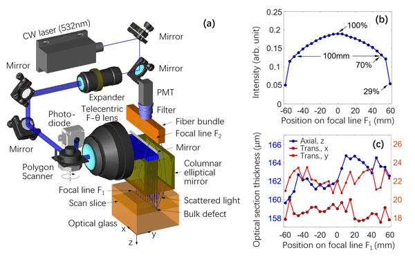
Fig. 1 (a) The dark-field line confocal imaging system. (b) The extended confocal line field. (c) The optical sectioning thicknesses in x, y and z directions and their uniformities within the confocal line field.