• Chinese Journal of Lasers
  • Vol. 51, Issue 3, 0307101 (2024)
Qiyu Bo1, Yuchen Wu1, Siqi Qiu4、5, and Zhiqing Zhang1、2、3、*
Author Affiliations
  • 1Institute of Modern Optics, Nankai University, Tianjin 300350, China
  • 2State Key Laboratory of Applied Optics, Changchun Institute of Optics, Fine Mechanics and Physics, Chinese Academy of Sciences, Changchun 130033, Jilin, China
  • 3Tianjin Key Laboratory of Micro-Scale Optical Information Science and Technology, Institute of Modern Optics, Nankai University, Tianjin 300350, China
  • 4Diagosis and Treatment Center of Breast Diseases, Shantou Central Hospital, Shantou 515041, Guangdong, China
  • 5Clinical Research Center, Shantou Central Hospital, Shantou 515041, Guangdong, China
  • show less
    DOI: 10.3788/CJL231383 Cite this Article Set citation alerts
    Qiyu Bo, Yuchen Wu, Siqi Qiu, Zhiqing Zhang. Current Progress of Third Harmonic Generation Microscopy in Tumor Diagnosis[J]. Chinese Journal of Lasers, 2024, 51(3): 0307101 Copy Citation Text show less
    Principle and experimental program for third harmonic generation (THG) microscopy. (a) Schematic diagram of THG and spontaneous fluorescence energy levels; (b) schematic diagram of the working bands of THG and multiphoton fluorescence signals; (c) a schematic showing whether THG signal can be emitted out in different parts of two media; (d) schematic diagram of THG microscopy experimental system (GS: galvo scanning; SL: scan lens; TL: tube lens; DM: dichroic mirror; OBJ: objective lens; IF: interference filter; L: lens)
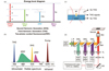
    Fig. 1. Principle and experimental program for third harmonic generation (THG) microscopy. (a) Schematic diagram of THG and spontaneous fluorescence energy levels; (b) schematic diagram of the working bands of THG and multiphoton fluorescence signals; (c) a schematic showing whether THG signal can be emitted out in different parts of two media; (d) schematic diagram of THG microscopy experimental system (GS: galvo scanning; SL: scan lens; TL: tube lens; DM: dichroic mirror; OBJ: objective lens; IF: interference filter; L: lens)
    Normal human brain tissue and gliomas images using THG (green) and SHG (red) imaging, the overlap between them is yellow. (a) Normal gray matter of the brain; (b) normal white matter of the brain; (c) low-grade diffuse gliomas (WHO grade II); (d) high-grade diffuse gliomas (WHO grade IV)
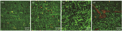
    Fig. 2. Normal human brain tissue and gliomas images using THG (green) and SHG (red) imaging, the overlap between them is yellow. (a) Normal gray matter of the brain; (b) normal white matter of the brain; (c) low-grade diffuse gliomas (WHO grade II); (d) high-grade diffuse gliomas (WHO grade IV)
    Human healthy lung tissue and lung cancer tissue were imaged with THG (green), SHG (red), and 2PEF (blue) signal, and the other colors in the pictures are superimposed on these three colors[51]. (a) Fresh healthy lung tissue with collagen, elastin and macrophages; (b) fresh epithelioid mesothelioma tissue, taken from bronchial biopsy, showed abnormal tumor cells and a small amount of collagen
    Fig. 3. Human healthy lung tissue and lung cancer tissue were imaged with THG (green), SHG (red), and 2PEF (blue) signal, and the other colors in the pictures are superimposed on these three colors[51]. (a) Fresh healthy lung tissue with collagen, elastin and macrophages; (b) fresh epithelioid mesothelioma tissue, taken from bronchial biopsy, showed abnormal tumor cells and a small amount of collagen
    Application example of compact portable intraoperative real-time diagnostic equipment. During the operation, a portion of tissue is resected, the upper cover is opened to place a sample into the instrument, and then THG/SHG/2PEF imaging is performed to diagnose whether the tissue is normal or tumor. If it were a tumor tissue, the type and the grade would be diagnosed. This can instruct the doctor to continue the operation (drawing from the portable compact higher harmonic microscopy system FD1070[51] developed by Professor Groot's team at VUA in the Netherlands)
    Fig. 4. Application example of compact portable intraoperative real-time diagnostic equipment. During the operation, a portion of tissue is resected, the upper cover is opened to place a sample into the instrument, and then THG/SHG/2PEF imaging is performed to diagnose whether the tissue is normal or tumor. If it were a tumor tissue, the type and the grade would be diagnosed. This can instruct the doctor to continue the operation (drawing from the portable compact higher harmonic microscopy system FD1070[51] developed by Professor Groot's team at VUA in the Netherlands)
    Schematic diagram of the endoscopic imaging principle of handheld fiber optic probes using nonlinear optical microscopy technology[68] (redrawn schematic diagram of a handheld fast fiber optic endoscope designed by the Li Xingde's team at Hopkins University in the United States in 2018)
    Fig. 5. Schematic diagram of the endoscopic imaging principle of handheld fiber optic probes using nonlinear optical microscopy technology[68] (redrawn schematic diagram of a handheld fast fiber optic endoscope designed by the Li Xingde's team at Hopkins University in the United States in 2018)
    Qiyu Bo, Yuchen Wu, Siqi Qiu, Zhiqing Zhang. Current Progress of Third Harmonic Generation Microscopy in Tumor Diagnosis[J]. Chinese Journal of Lasers, 2024, 51(3): 0307101
    Download Citation