• Laser & Optoelectronics Progress
  • Vol. 58, Issue 18, 1811023 (2021)
Ruijuan Gao, Chunhua Wang*, Jinxing Ning, Keshuai Wu, and Xiang Gu
Author Affiliations
  • Key Laboratory of Specialty Fiber Optics and Optical Access Networks, Shanghai University, Shanghai 200444, China
  • show less
    DOI: 10.3788/LOP202158.1811023 Cite this Article Set citation alerts
    Ruijuan Gao, Chunhua Wang, Jinxing Ning, Keshuai Wu, Xiang Gu. Polarization Microscopy Imaging of Biological Cells Based on Mueller Matrix[J]. Laser & Optoelectronics Progress, 2021, 58(18): 1811023 Copy Citation Text show less
    Principle of the Mueller matrix polarization microscopy imaging system
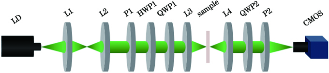
    Fig. 1. Principle of the Mueller matrix polarization microscopy imaging system
    Four uncorrelated PSG polarization states
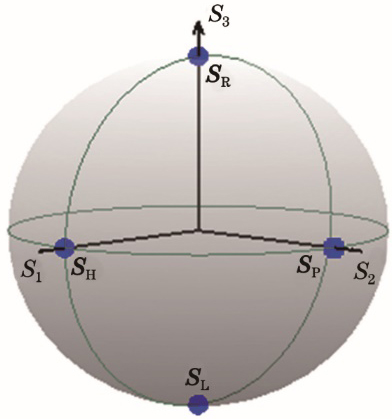
    Fig. 2. Four uncorrelated PSG polarization states
    Measured and theoretical values of the Mueller matrix
    Fig. 3. Measured and theoretical values of the Mueller matrix
    Normalized Mueller matrix imaging results of the inner epidermal tissue of onion. (a) Mass fraction is 0%; (b) mass fraction is 2.5%; (c) mass fraction is 5%; (d) mass fraction is 10%
    Fig. 4. Normalized Mueller matrix imaging results of the inner epidermal tissue of onion. (a) Mass fraction is 0%; (b) mass fraction is 2.5%; (c) mass fraction is 5%; (d) mass fraction is 10%
    Mueller matrix elements of onion inner epidermal tissue under different mass fractions of glucose solutions. (a) m22; (b) m23
    Fig. 5. Mueller matrix elements of onion inner epidermal tissue under different mass fractions of glucose solutions. (a) m22; (b) m23
    Mueller matrix polarization decomposition parameters of onion inner epidermal tissue in glucose solutions with different mass fractions. (a) Intensity; (b) depolarization; (c) circular retardance; (d) diattenuation
    Fig. 6. Mueller matrix polarization decomposition parameters of onion inner epidermal tissue in glucose solutions with different mass fractions. (a) Intensity; (b) depolarization; (c) circular retardance; (d) diattenuation
    Imaging results of human laryngeal carcinoma cells at different experimental time. (a) Intensity imaging; (b) Mueller matrice
    Fig. 7. Imaging results of human laryngeal carcinoma cells at different experimental time. (a) Intensity imaging; (b) Mueller matrice
    Mueller matrix polarization decomposition imaging of human laryngeal carcinoma cells at different experimental time. (a) Depolarization; (b) circular retardance; (c) diattenuation
    Fig. 8. Mueller matrix polarization decomposition imaging of human laryngeal carcinoma cells at different experimental time. (a) Depolarization; (b) circular retardance; (c) diattenuation
    Polarization statePSGPSA
    P1HWP1QWP1QWP2P2
    SH00000
    SL00-45045
    SR00454545
    SP022.5459045
    Table 1. Collimation direction of the optical device unit: (°)
    ElementFormula
    m11ILL+ILP+IRL+IRP
    m12-ILL-ILP-IRL-IRP+2IHL+2IHP
    m13-ILL-ILP-IRL-IRP+2IPL+2IPP
    m14-ILL-ILP+IRL+IRP
    m21-ILL-ILP-IRL-IRP+2ILH+2IRH
    m22ILL+ILR+IRL+IRP-2ILH-2IRH-2IHL-2IHP+4IHH
    m23ILL+ILP+IRL+IRP-2ILH-2IRH-2IPL-2IPP-4IPH
    m24ILL+ILP-IRL-IRP-2ILH+2IRH
    m31-ILL-ILP-IRL-IRP+2ILR+2IRR
    m32ILL+ILP+IRL+IRP-2ILR-2IRR-2IHL-2IHP+4IHR
    m33ILL+ILP+IRL-IRP-2ILR-2IRR-2IPL-2IPP+4IPR
    m34ILL+ILP-IRL-IRP-2ILR+2IRR
    m41ILL-ILP+IRL-IRP
    m42-ILL+ILP-IRL+IRP+2IHL-2IHP
    m43-ILL+ILP-IRL+IRP+2IPL-2IPP
    m44-ILL+ILP+IRL-IRP
    Table 2. Calculation formulae of the Mueller matrix elements
    Ruijuan Gao, Chunhua Wang, Jinxing Ning, Keshuai Wu, Xiang Gu. Polarization Microscopy Imaging of Biological Cells Based on Mueller Matrix[J]. Laser & Optoelectronics Progress, 2021, 58(18): 1811023
    Download Citation