
Fuming Xie, Ni Yao, Wei Fang, Haifeng Wang, Fuxing Gu, Songlin Zhuang. Single-mode lasing via loss engineering in fiber-taper-coupled polymer bottle microresonators[J]. Photonics Research, 2017, 5(6): B29
- Photonics Research
- Vol. 5, Issue 6, B29 (2017)
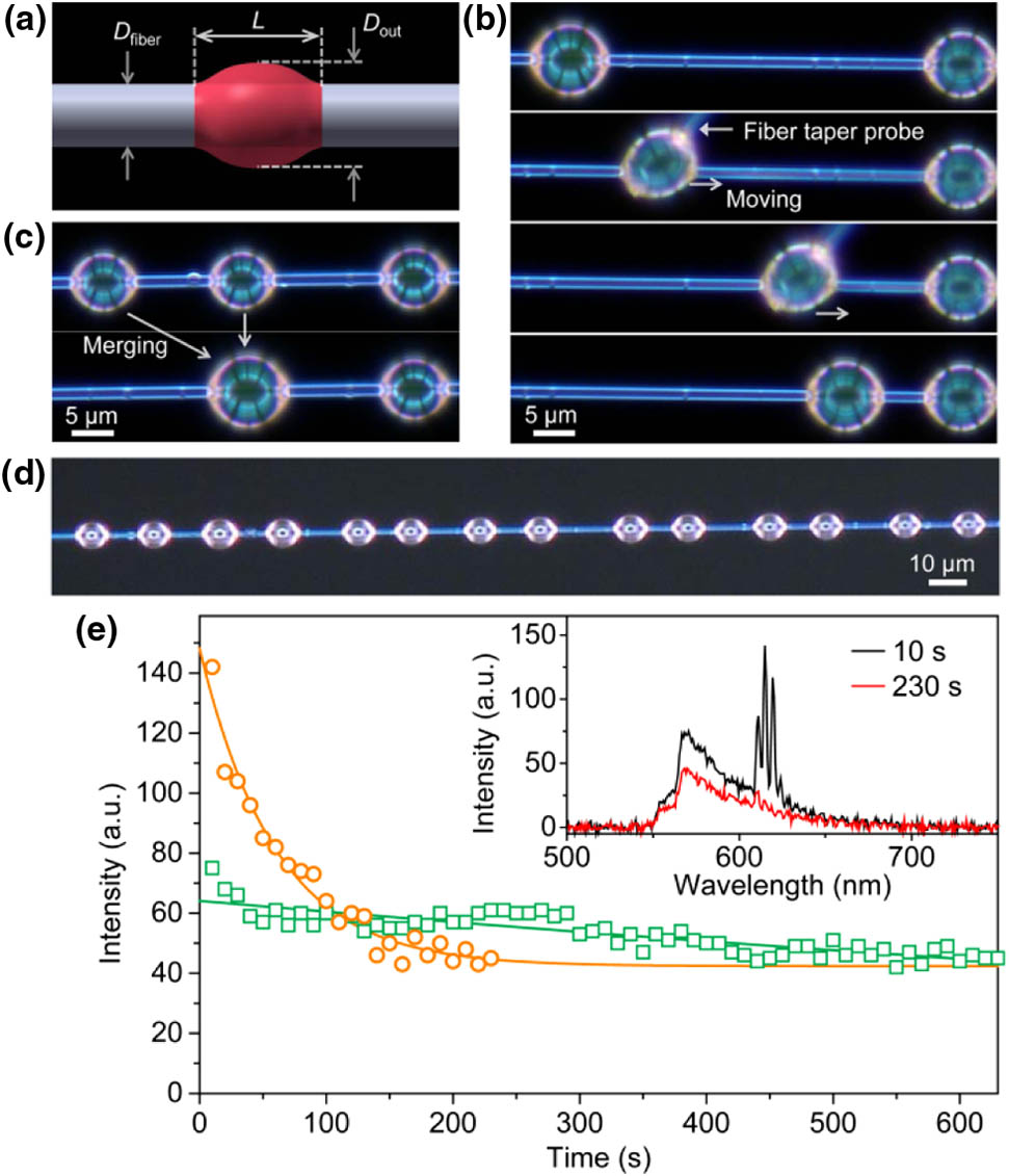
Fig. 1. (a) Definition of D out D fiber L
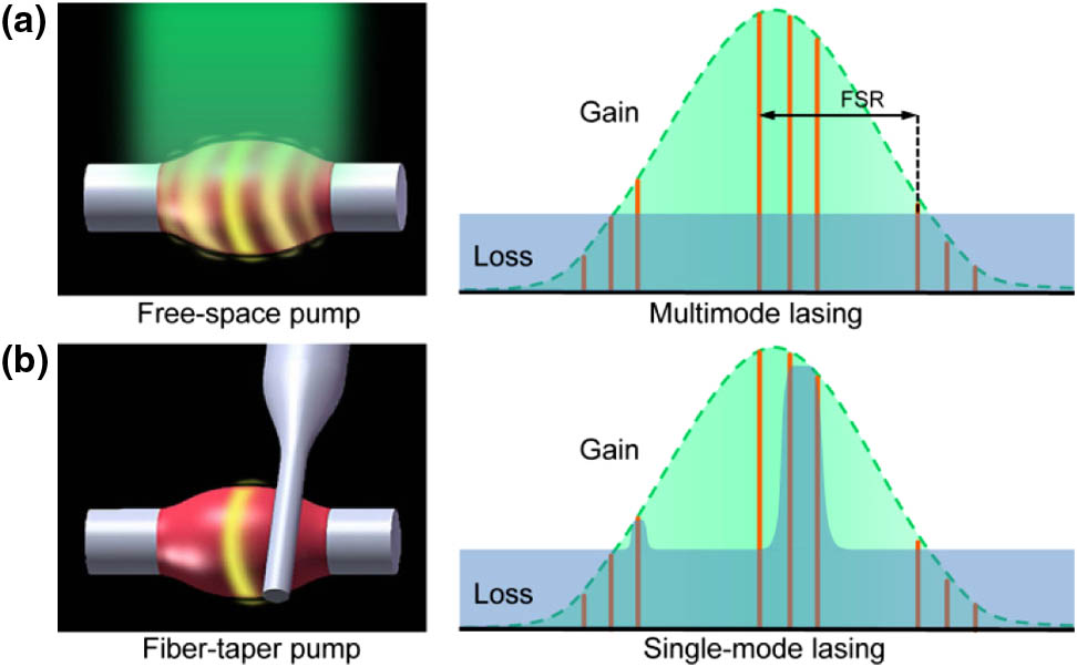
Fig. 2. Principle of single WGM lasing in a polymer bottle microresonator. (a) Multimode lasing behavior under uniform pump. (b) Single WGM lasing by adjusting the coupling position to suppress high-order modes.
Fig. 3. (a) Illustration of fiber-taper-coupled bottle microresonator. Definition of D FT D out = 5.5 μm D fiber = 3.2 μm L = 7.5 μm P in = 26.1 nJ
Fig. 4. (a) Lasing spectra and (b) their corresponding microscope images of a polymer bottle microresonator (D out = 6.1 μm D fiber = 3.9 μm L = 10.8 μm D out = 5.3 μm D fiber = 3.9 μm L = 7.6 μm
Fig. 5. Electric field-intensity distributions of (a) TM 43 1 TM 42 2 TM 42 3 TM 41 4
Fig. 6. (a) Lasing spectra and (b) their corresponding microscope images of a polymer bottle microresonator (D out = 6.1 μm D fiber = 3.9 μm L = 10.8 μm D out = 5.3 μm D fiber = 3.9 μm L = 7.6 μm

Set citation alerts for the article
Please enter your email address
© Copyright 2018-2021 | Chinese Laser Press. All Rights Reserved 沪ICP备15018463号-20