• Chinese Optics Letters
  • Vol. 15, Issue 5, 051701 (2017)
Hanh N. D. Le1、*, Ryan Decker2, Axel Krieger2, and Jin U. Kang1
Author Affiliations
  • 1Electrical and Computer Engineering Department, Johns Hopkins University, Baltimore, MD 21218, USA
  • 2Sheikh Zayed Institute for Pediatric Surgical Innovation, Children’s National Health System, Washington, DC 20010, USA
  • show less
    DOI: 10.3788/COL201715.051701 Cite this Article Set citation alerts
    Hanh N. D. Le, Ryan Decker, Axel Krieger, Jin U. Kang. Experimental assessment of a 3-D plenoptic endoscopic imaging system[J]. Chinese Optics Letters, 2017, 15(5): 051701 Copy Citation Text show less
    Schematic of the endoscopic setup with the plenoptic camera and relay lens system.
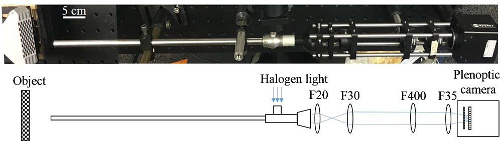
    Fig. 1. Schematic of the endoscopic setup with the plenoptic camera and relay lens system.
    Triangulation principle for virtual depth estimation. IP: Image plane.
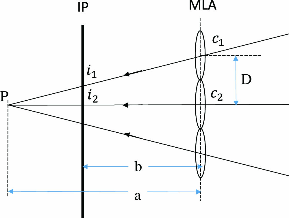
    Fig. 2. Triangulation principle for virtual depth estimation. IP: Image plane.
    Resolution and contrast measurements.
    Fig. 3. Resolution and contrast measurements.
    (a) Microlens image of the checker board at 0° with (b) its depth map and (c, d) point cloud data at different views.
    Fig. 4. (a) Microlens image of the checker board at 0° with (b) its depth map and (c, d) point cloud data at different views.
    Microlens image of the checker board at 45° with (b) its depth map and (c, d) point cloud data at different views.
    Fig. 5. Microlens image of the checker board at 45° with (b) its depth map and (c, d) point cloud data at different views.
    (a–c) Microlens image of a plane and inhomogeneous objects and (d–f) its reconstructed depth maps.
    Fig. 6. (a–c) Microlens image of a plane and inhomogeneous objects and (d–f) its reconstructed depth maps.
    (a) Microlens image of a fowl ventricular specimen and (b–d) its 3-D reconstructions at multiple angles.
    Fig. 7. (a) Microlens image of a fowl ventricular specimen and (b–d) its 3-D reconstructions at multiple angles.
      45°
    AccuracyMean0.0850.818
    Standard deviation0.0320.440
    Maximum0.1031.439
    PrecisionMean1.1412.367
    Standard deviation0.7211.800
    Maximum3.86311.658
    Table 1. Reconstruction Accuracy and Precision at Two Planar Angle Deviations at 0° and 45° (Unit: mm)
    Hanh N. D. Le, Ryan Decker, Axel Krieger, Jin U. Kang. Experimental assessment of a 3-D plenoptic endoscopic imaging system[J]. Chinese Optics Letters, 2017, 15(5): 051701
    Download Citation