• Advanced Photonics
  • Vol. 6, Issue 2, 026004 (2024)
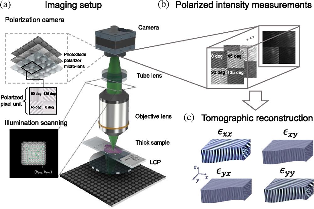
Shiqi Xu1, Xi Yang1, Paul Ritter2, Xiang Dai1、3, Kyung Chul Lee1、4, Lucas Kreiss1、2, Kevin C. Zhou1、5, Kanghyun Kim1, Amey Chaware1, Jadee Neff6, Carolyn Glass6, Seung Ah Lee4, Oliver Friedrich2, and Roarke Horstmeyer1、*
Author Affiliations
  • 1Duke University, Durham, North Carolina, United States
  • 2Friedrich-Alexander University, Erlangen, Germany
  • 3UC San Diego, La Jolla, California, United States
  • 4Yonsei University, Seoul, Republic of Korea
  • 5UC Berkeley, Berkeley, California, United States
  • 6Duke University Medical Center, Durham, North Carolina, United States
  • show less
    DOI: 10.1117/1.AP.6.2.026004 Cite this Article Set citation alerts
    Shiqi Xu, Xi Yang, Paul Ritter, Xiang Dai, Kyung Chul Lee, Lucas Kreiss, Kevin C. Zhou, Kanghyun Kim, Amey Chaware, Jadee Neff, Carolyn Glass, Seung Ah Lee, Oliver Friedrich, Roarke Horstmeyer. Tensorial tomographic Fourier ptychography with applications to muscle tissue imaging[J]. Advanced Photonics, 2024, 6(2): 026004 Copy Citation Text show less

    Abstract

    We report tensorial tomographic Fourier ptychography (T2oFu), a nonscanning label-free tomographic microscopy method for simultaneous imaging of quantitative phase and anisotropic specimen information in 3D. Built upon Fourier ptychography, a quantitative phase imaging technique, T2oFu additionally highlights the vectorial nature of light. The imaging setup consists of a standard microscope equipped with an LED matrix, a polarization generator, and a polarization-sensitive camera. Permittivity tensors of anisotropic samples are computationally recovered from polarized intensity measurements across three dimensions. We demonstrate T2oFu’s efficiency through volumetric reconstructions of refractive index, birefringence, and orientation for various validation samples, as well as tissue samples from muscle fibers and diseased heart tissue. Our reconstructions of healthy muscle fibers reveal their 3D fine-filament structures with consistent orientations. Additionally, we demonstrate reconstructions of a heart tissue sample that carries important polarization information for detecting cardiac amyloidosis.
    Supplementary Materials
    Shiqi Xu, Xi Yang, Paul Ritter, Xiang Dai, Kyung Chul Lee, Lucas Kreiss, Kevin C. Zhou, Kanghyun Kim, Amey Chaware, Jadee Neff, Carolyn Glass, Seung Ah Lee, Oliver Friedrich, Roarke Horstmeyer. Tensorial tomographic Fourier ptychography with applications to muscle tissue imaging[J]. Advanced Photonics, 2024, 6(2): 026004
    Download Citation