• Spectroscopy and Spectral Analysis
  • Vol. 40, Issue 10, 3021 (2020)
Hui-yan TIAN1、1、*, Yu LIU1、1, Jiao-qi HUANG1、1, Feng-xin XIE1、1, Guo-rong HUANG1、1, Pu LIAO1、1, Wei-ling FU1、1, and Yang ZHANG1、1
Author Affiliations
  • 1[in Chinese]
  • 11. Department of Clinical Laboratory, The First Affiliated Hospital of the Army Military Medical University, Chongqing 400038, China
  • show less
    DOI: 10.3964/j.issn.1000-0593(2020)10-3021-08 Cite this Article
    Hui-yan TIAN, Yu LIU, Jiao-qi HUANG, Feng-xin XIE, Guo-rong HUANG, Pu LIAO, Wei-ling FU, Yang ZHANG. Research Progress and Application of Surface-Enhanced Raman Scattering Technique in Nucleic Acid Detection[J]. Spectroscopy and Spectral Analysis, 2020, 40(10): 3021 Copy Citation Text show less
    Principle of SERS(a) Illustration of the collective oscillation of free electrons in metal nanoparticles upon excitation by an electromagnetic wave; (b) Chemical enhancement results from charge transfer resonance between signal molecules and metal nanostructures, which is usually weaker than electromagnetic enhancement
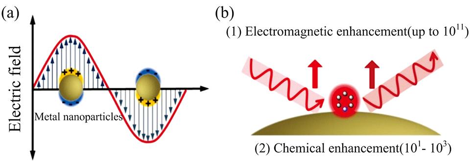
    Fig. 1. Principle of SERS
    (a) Illustration of the collective oscillation of free electrons in metal nanoparticles upon excitation by an electromagnetic wave; (b) Chemical enhancement results from charge transfer resonance between signal molecules and metal nanostructures, which is usually weaker than electromagnetic enhancement
    SERS-based nucleic acid analysis using a label-free SERS approach (left) or SERS tags (right)In label-free SERS, the spectroscopic signal results from analyte adsorption onto the SERS substrate, whereas in SERS tags-based specific recognition assays, the spectroscopic signal results from the reporter molecules on the SERS tags
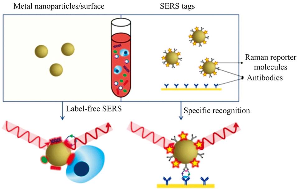
    Fig. 2. SERS-based nucleic acid analysis using a label-free SERS approach (left) or SERS tags (right)
    In label-free SERS, the spectroscopic signal results from analyte adsorption onto the SERS substrate, whereas in SERS tags-based specific recognition assays, the spectroscopic signal results from the reporter molecules on the SERS tags
    Scheme of SERS-based nucleic acid detection based on sandwich structure[18]
    Fig. 3. Scheme of SERS-based nucleic acid detection based on sandwich structure[18]
    (A) Schematic illustration of the synthetic procedures of Raman dye-coded Au-RNNPs using DNA-modified AuNPs as templates (a) and Ag-HMSs using bacteria as template; (B) Schematic illustration of the multiplex SERS assay for triple-target miRNA detection; (C)SERS spectra of the nanoprobes obtained in the presence of (a) single and (b) multiple miRNA[19]
    Fig. 4. (A) Schematic illustration of the synthetic procedures of Raman dye-coded Au-RNNPs using DNA-modified AuNPs as templates (a) and Ag-HMSs using bacteria as template; (B) Schematic illustration of the multiplex SERS assay for triple-target miRNA detection; (C)SERS spectra of the nanoprobes obtained in the presence of (a) single and (b) multiple miRNA[19]
    Scheme of SERS-based nucleic acid detection based on hairpin structure[18]
    Fig. 5. Scheme of SERS-based nucleic acid detection based on hairpin structure[18]
    (a) Schematic illustration of the molecular beacons functionalized-SERS sensor for simultaneously measuring multiple miRNAs[24]; (b) Detection scheme of the SERS “inverse Molecular Sentinel” nanoprobes[25]
    Fig. 6. (a) Schematic illustration of the molecular beacons functionalized-SERS sensor for simultaneously measuring multiple miRNAs[24]; (b) Detection scheme of the SERS “inverse Molecular Sentinel” nanoprobes[25]
    Scheme of SERS-based nucleic acid detection based on the signal amplification of hybridization chain reaction[26,27]
    Fig. 7. Scheme of SERS-based nucleic acid detection based on the signal amplification of hybridization chain reaction[26,27]
    检测方法优点缺点
    无标记检测(1) 步骤简单, 无需对样品进行处理, 可以避免对生物体的破坏, 最大限度地保持生物体的活性;
    (2)可以充分发挥SERS作为振动光谱具有指纹识别的优势, 得到DNA自身的结构信息。
    (1) 在实际SERS检测中, 由于碱基的拉曼散射截面很小, 导致DNA/RNA的SERS信号很弱, 检测灵敏度较低;
    (2) 不同的DNA/RNA序列具有相同的碱基种类只是碱基排列顺序不同, 因而具有相似的SERS光谱, 很难直接通过SERS信号来确定特定的DNA序列, 需要借助一些数据分析手段。
    标记型检测特异性好, 灵敏度高, 可直接进行定量检测在检测结构及探针的设计上有一定的难度。
    Table 1. Analysis of the label-free SERS approach and SERS tags-based nucleic acid assays
    目标核酸SERS检测方法LOD/判别正确率参考文献
    肝癌miRNA标志物(miRNA-21, miRNA-122, miRNA-223)“夹心法”检测: Ag微球偶联DNA捕获链, Au RNNP-44DP/4ATP/DTNB DNA探针10 fmol·L-1[15]
    乙型肝炎病毒DNA“夹心法”检测: Ag nanorice @MGITC @SiO250 amol·L-1[16]
    复合病原体 DNA“夹心法”检测: Au NPS DNA探针, 150 nm 的金纳米棒上构建检测基底10 pmol·L-1[17]
    肺癌miRNA标志物(miRNA 21, miRNA 486, miRNA 375)“信号开—关法”: 发夹探针-ROX/Cy5/FAM, Ag NRs微阵列增强基底miRNA 21/393 amol·L-1, miRNA 486/176 amol·L-1, miRNA 375/144 amol·L-1[20]
    肺癌miRNA标志物(miRNA 21, miRNA 34a)“信号关—开法”: 发夹探针-Cy5/Cy5.5, Ag-coated Au nanostars增强基底成功实现对生物样本中混合miRNA标志物的检测[21]
    miRNA 141“HCR信号放大法”: Au NPS在DNA聚合物上形成“热点”效应0.17 fmol·L-1[22]
    miRNA 21“HCR信号放大法”: Ag NPS在DNA聚合物上形成“热点”效应对单碱基突变及双碱基突变序列的检测信号点分别仅为12.4%和4.6%(定义对靶序列miRNA-21的检测信号点100%)[23]
    miRNA-150“信号开—关法”: 发夹探针-Cy5-Au NPS70.2 amol·L-1[24]
    DNA“热点效应”: 通过ss-DNA的自组装在Au NPS间形成“热点”, 再利用酶切技术进行“热点”消除通过检测拉曼报告分子拉曼信号的变化, 证明该方法可行[25]
    Table 2. Typical research about SERS used for detection on nucleic acids
    Hui-yan TIAN, Yu LIU, Jiao-qi HUANG, Feng-xin XIE, Guo-rong HUANG, Pu LIAO, Wei-ling FU, Yang ZHANG. Research Progress and Application of Surface-Enhanced Raman Scattering Technique in Nucleic Acid Detection[J]. Spectroscopy and Spectral Analysis, 2020, 40(10): 3021
    Download Citation