
Yao Tong, Qin Wang, Heng Yang, Xiaoting Liu, Enrou Mei, Xiaojuan Liang, Zhijun Zhang, Weidong Xiang, "Enhanced multimodal luminescence and ultrahigh stability Eu3+-doped CsPbBr3 glasses for X-ray detection and imaging," Photonics Res. 9, 2369 (2021)
- Photonics Research
- Vol. 9, Issue 12, 2369 (2021)
Abstract
1. INTRODUCTION
As a category of distinctive luminescent materials, the scintillator is in a position to convert high-energy photon (X-ray, and
In recent years, perovskite materials, especially all-inorganic halide perovskites (
The glass matrix is identified as a superior compactness carrier that protects NCs from environmental damage, and simultaneously maintains excellent optical properties [27–29]. However, the uniformity and luminescence properties of perovskite glass also hinder its application in X-ray imaging. Herein, we synthesized
Sign up for Photonics Research TOC. Get the latest issue of Photonics Research delivered right to you!Sign up now
2. EXPERIMENT
A. Chemicals
Boron oxide (
B. Synthesis of CPB-xEu
A series of borosilicate CPB-xEu was synthesized with the arranged molar proportions of 75% (
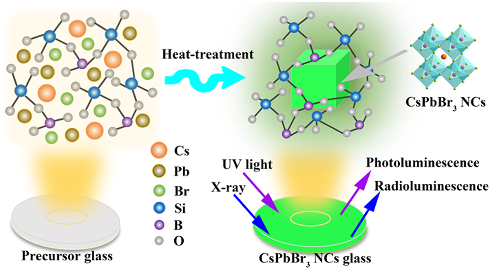
Figure 1.Schematic illustration of the
C. Structural Characterization
The crystal structure of the sample was determined by an X-ray diffractometer (Bruker, D8 Advance) with Cu
3. RESULTS AND DISCUSSION
To obtain the optimal optical properties of CPB-0Eu glass, the precursor glasses were heat-treated at 480°C–540°C for 10 h. As shown in Fig. 2(a), the diffraction peak is not obvious and shows only an amorphous halo when the heat-treatment temperature is 480°C for 10 h. With the rise of heat-treatment temperature, X-ray diffraction (XRD) peaks appear by degrees and show a slight redshift, but always correspond well to
![]()
Figure 2.(a) XRD patterns of CPB-0Eu at different heat-treatment temperatures. (b) TEM image of CPB-0Eu at 520°C for 10 h. (c) PL spectra under UV light excitation of CPB-0Eu at different heat-treatment temperatures. (d) HRTEM image of CPB-0Eu at 520°C for 10 h. (e) Standard structure model diagram of
Subsequently, a series of
![]()
Figure 3.(a) Images of CPB-
Similarly, to better verify the microstructure changes of
![]()
Figure 4.(a) TEM image of CPB-1.8Eu (illustration is the HRTEM image of CPB-1.8Eu at 520°C for 10 h). (b) XPS survey spectra of CPB-0Eu and CPB-1.8Eu. (c) EDX mapping images of CPB-1.8Eu. Corresponding high-resolution spectra of (d) Cs 3d for CPB-0Eu and CPB-1.8Eu, (e) Pb 4f for CPB-
![]()
Figure 5.TEM images and histograms of size distribution of (a) CPB-0Eu and (b) CPB-1.8Eu. (c) Transmittance spectra of CPB-
To evaluate the potential scintillation properties of CPB-xEu, the RL spectra of CPB-xEu (
![]()
Figure 6.RL spectra of (a) CPB-0Eu, (b) CPB-0.6Eu, and (c) CPB-1.8Eu upon increasing current of X-ray cathode tube. (d), (e) Schematic illustration of RL mechanism about CPB-
Based on the unique scintillation property of
Subsequently, we compared and investigated the change of integrated radiation intensity dependence on the time of CPB-0.6Eu and CPB-0Eu glasses at a radiation dose of
Benefiting from the optimized luminescence properties, enhanced uniformity, and attenuated scattering of the CPB-xEu (
![]()
Figure 7.(a) Schematic of the experimental setup used for X-ray imaging with sample (X-rays are excited directly at the side of the sample; voltage: 50 kV, dose rate:
4. CONCLUSION
In summary, a series of enhanced multimodal luminescence CPB-xEu glasses with prominent PLQY and good uniformity was acquired by conventional high-temperature sintering and subsequent heat treatment. Surprisingly, the emission peak of the fabricated CPB-xEu glasses appeared close to 528 nm and maintained excellent luminescence. We confirmed the successful incorporation of
APPENDIX A
Figure
![]()
Figure 8.Relative PL intensity at five different locations of the same sample.
![]()
Figure 9.(a) Excitation spectra of CPB-
![]()
Figure 10.Characteristic PL spectra of Eu ions at 560–700 nm under different wavelength excitation of CPB-0.6Eu.
![]()
Figure 11.PL decay of CPB-0Eu and CPB-0.6Eu glasses.
![]()
Figure 12.RL spectra of CPB-
![]()
Figure 13.Linear relation based on RL intensity and X-ray dose rate.
![]()
Figure 14.The relative RL intensity of CPB-0.6Eu and CPB-0Eu glass depending on time of X-ray radiation at a radiation dose of 105 μGyair/s and a voltage of 50 kV. The illustration shows a histogram of Pb2+ solubility after 5 days of water immersion before and after X-ray radiation.
References
[1] A. Horimoto, N. Kawano, D. Nakauchi, H. Kimura, M. Akatsuka, T. Yanagida. Scintillation properties of organic–inorganic perovskite-type compounds with fluorophenethylamine. Opt. Mater., 101, 109686(2020).
[2] D. Yu, P. Wang, F. Cao, Y. Gu, J. Liu, Z. Han, B. Huang, Y. Zou, X. Xu, H. Zeng. Two-dimensional halide perovskite as β-rayscintillator for nuclear radiation monitoring. Nat. Commun., 11, 3395(2020).
[3] F. Maddalena, L. Tjahjana, A. Xie, , S. Zeng, H. Wang, P. Coquet, W. Drozdowski, C. Dujardin, C. Dang, M. Birowosuto. Inorganic, organic, and perovskite halides with nanotechnology for high–light yield X- and γ-ray scintillators. Crystals, 9, 88(2019).
[4] K. S. Kim, S. Y. Kang, W. S. Kim, H. S. Cho, C. K. Park, D. Y. Lee, G. A. Kim, S. Y. Park, H. W. Lim, H. W. Lee, J. E. Park, D. H. Jeon, Y. H. Lim, U. K. Je, T. H. Woo. Improvement of radiographic visibility using an image restoration method based on a simple radiographic scattering model for X-ray nondestructive testing. NDT & E Int., 98, 117-122(2018).
[5] P. Lecoq. Development of new scintillators for medical applications. Nucl. Instrum. Methods Phys. Res. A, 809, 130-139(2016).
[6] H. Wei, J. Huang. Halide lead perovskites for ionizing radiation detection. Nat. Commun., 10, 1066(2019).
[7] A. Phunpueok, V. Thongpool, S. Jaiyen. Scintillation response of CdWO4 scintillator for gamma-ray detection. J. Phys. Conf. Ser., 1380, 012128(2019).
[8] M. Tyagi, A. Khan, P. Q. Vuong, H. J. Kim. Comprehending the role of trap centers and host energy transfers in excitation density dependent kinetics of Ce doped Gd3Ga3Al2O12 scintillator; an unresolved scintillation characteristic. J. Lumin., 219, 116815(2020).
[9] Y. Wu, J. Peng, D. Rutstrom, M. Koschan, C. Foster, C. L. Melcher. Unraveling the critical role of site occupancy of lithium codopants in Lu2SiO5:Ce3+ single-crystalline scintillators. ACS. Appl. Mater. Interfaces, 11, 8194-8201(2019).
[10] A. Khan, D. J. Daniel, M. Tyagi, H. J. Kim, M. H. Lee, Y. Kim. Czochralski growth, electronic structure, luminescence and scintillation properties of Cs2Mo3O10: a new scintillation crystal for ovββ decay search. J. Alloys Comp., 821, 153466(2020).
[11] K. Kagami, M. Koshimizu, Y. Fujimoto, S. Kishimoto, R. Haruki, F. Nishikido, K. Asai. High-energy X-ray detection capabilities of Hf-loaded plastic scintillators synthesized by sol–gel method. J. Mater. Sci., 31, 896-902(2019).
[12] B. K. Cha, J. Y. Kim, T. J. Kim, C. Sim, G. Cho. Fabrication and imaging characterization of high sensitive CsI(Tl) and Gd2O2S(Tb) scintillator screens for X-ray imaging detectors. Radiat. Meas., 45, 742-745(2010).
[13] O. Guillot-Noël, J. T. M. de Haas, P. Dorenbos, C. W. E. van Eijk, K. Krämer, H. U. Güdel. Optical and scintillation properties of cerium-doped LaCl3, LuBr3 and LuCl3. J. Lumin., 85, 21-35(1999).
[14] A. S. Pushak, V. V. Vistovskyy, T. M. Demkiv, A. A. Halyatkin, Y. R. Datsyuk, A. V. Gektin, S. V. Myagkota, P. A. Rodnyi, A. S. Voloshinovskii. Luminescence of SrX2 microcrystals (X = Cl, I) activated with Eu2+ ions and dispersed in a NaI matrix. Opt. Spectrosc., 117, 593-597(2014).
[15] J. M. Ogieglo, A. Katelnikovas, A. Zych, T. Justel, A. Meijerink, C. R. Ronda. Luminescence and luminescence quenching in Gd3(Ga, Al)5O12 scintillators doped with Ce3+. J. Phys. Chem. A., 117, 2479-2484(2013).
[16] M. Mori, J. Xu, G. Okada, T. Yanagida, J. Ueda, S. Tanabe. Comparative study of optical and scintillation properties of Ce:YAGG, Ce:GAGG and Ce:LuAGG transparent ceramics. J. Ceram. Soc. Jpn., 124, 569-573(2016).
[17] A. Xie, T. H. Nguyen, C. Hettiarachchi, M. E. Witkowski, W. Drozdowski, M. D. Birowosuto, H. Wang, C. Dang. Thermal quenching and dose studies of X-ray luminescence in single crystals of halide perovskites. J. Phys. Chem. C, 122, 16265(2018).
[18] C. Ji, S. Wang, Y. Wang, H. Chen, L. Li, Z. Sun, Y. Sui, S. Wang, J. Luo. 2D hybrid perovskite ferroelectric enables highly sensitive X-ray detection with low driving voltage. Adv. Funct. Mater., 30, 1905529(2019).
[19] W. Pan, B. Yang, G. Niu, K. H. Xue, X. Du, L. Yin, M. Zhang, H. Wu, X. S. Miao, J. Tang. Hot-pressed CsPbBr3 quasi-monocrystalline film for sensitive direct X-ray detection. Adv. Mater., 31, 1904405(2019).
[20] F. R. Hu, H. C. Zhang, C. Sun, C. Y. Yin, B. H. Lv, C. F. Zhang, W. W. Yu, X. Y. Wang, Y. Zhang, M. Xiao. Superior optical properties of perovskite nanocrystals as single photon emitters. ACS. Nano., 9, 12410-12416(2015).
[21] J. H. Heo, D. H. Shin, J. K. Park, D. H. Kim, S. J. Lee, S. H. Im. High-performance next-generation perovskite nanocrystal scintillator for nondestructive X-ray imaging. Adv. Mater., 30, 1801743(2018).
[22] Q. Chen, J. Wu, X. Ou, B. Huang, J. Almutlaq, A. A. Zhumekenov, X. Guan, S. Han, L. Liang, Z. Yi, J. Li, X. Xie, Y. Wang, Y. Li, D. Fan, D. B. L. Teh, A. H. All, O. F. Mohammed, O. M. Bakr, T. Wu, M. Bettinelli, H. Yang, W. Huang, X. Liu. All-inorganic perovskite nanocrystal scintillators. Nature, 561, 88-93(2018).
[23] C. C. Stoumpos, C. D. Malliakas, J. A. Peters, Z. Liu, M. Sebastian, J. Im, T. C. Chasapis, A. C. Wibowo, D. Y. Chung, A. J. Freeman, B. W. Wessels, M. G. Kanatzidis. Crystal growth of the perovskite semiconductor CsPbBr3: a new material for high-energy radiation detection. Cryst. Growth Des., 13, 2722-2727(2013).
[24] C. Zheng, C. Bi, F. Huang, D. Binks, J. Tian. Stable and strong emission CsPbBr3 quantum dots by surface engineering for high-performance optoelectronic films. ACS Appl. Mater. Interfaces, 11, 25410-25416(2019).
[25] F. Cao, D. Yu, W. Ma, X. Xu, B. Cai, Y. M. Yang, S. Liu, L. He, Y. Ke, S. Lan, K. L. Choy, H. Zeng. Shining emitter in a stable host: design of halide perovskite scintillators for X-ray imaging from commercial concept. ACS Nano, 14, 5183-5193(2020).
[26] C. Wang, H. Lin, Z. Zhang, Z. Qiu, H. Yang, Y. Cheng, J. Xu, X. Xiang, L. Zhang, Y. Wang. X-ray excited CsPb(Cl,Br)3 perovskite quantum dots-glass composite with long-lifetime. J. Eur. Ceram. Soc., 40, 2234-2238(2020).
[27] B. Zhang, K. Zhang, L. Li, C. Xu, R. Wang, C. Wang, J. Yang, Y. Yang, J. Wang, F. Qiu, T. Sun, C. Zhou, X. Wen. Enhancing stability and luminescence quantum yield of CsPbBr3 quantum dots by embedded in borosilicate glass. J. Alloy. Compd., 874, 159962(2021).
[28] P. Aryal, H. Kima, S. Saha, J. Cho, A. V. Ntarisa, S. Kothan, J. Kaewkhao. Rapid and convenient crystallization of quantum dot CsPbBr3 inside a phosphate glass matrix. J. Alloy. Compd., 866, 158974(2021).
[29] Y. Lu, P. Li, W. Xie, Y. Duan, X. Wang, S. Xu, J. Zhang. Negative thermal quenching CsPbBr3 glass-ceramic based on intrinsic radiation and vacancy defect co-induced dual-emission. J. Eur. Ceram. Soc., 41, 3635-3642(2021).
[30] Y. Ye, W. Zhang, Z. Zhao, J. Wang, C. Liu, Z. Deng, X. Zhao, J. Han. Highly luminescent cesium lead halide perovskite nanocrystals stabilized in glasses for light-emitting applications. Adv. Opt. Mater., 7, 1801663(2019).
[31] C. Shen, Y. Zhao, L. Yuan, L. Ding, Y. Chen, H. Yang, S. Liu, J. Nie, W. Xiang, X. Liang. Transition metal ion doping perovskite nanocrystals for high luminescence quantum yield. Chem. Eng. J., 382, 122868(2020).
[32] C. Daqin, F. Gaoliang, C. Xiao, L. Lei, Z. Jiasong, M. Qinan, Z. Su, L. Junni. Mn Doped CsPbCl3 perovskite nanocrystals: solvothermal synthesis, dual-color luminescence and improved stability. J. Phys. Chem. C, 6, 8990-8998(2018).
[33] C. M. Guvenc, Y. Yalcinkaya, S. Ozen, H. Sahin, M. M. Demir. Gd3+-doped α-CsPbI3 nanocrystals with better phase stability and optical properties. J. Phys. Chem. C, 123, 24865-24872(2019).
[34] P. Li, Y. Duan, Y. Lu, A. Xiao, Z. Zeng, S. Xu, J. Zhang. Nanocrystalline structure control and tunable luminescence mechanism of Eu-doped CsPbBr3 quantum dot glass for WLEDs. Nanoscale, 12, 6630-6636(2020).
[35] C. Wu, S. Zeng, Z. Wang, F. Wang, H. Zhou, J. Zhang, Z. Ci, L. Sun. Efficient mechanoluminescent elastomers for dual-responsive anticounterfeiting device and stretching/strain sensor with multimode sensibility. Adv. Funct. Mater., 28, 1803168(2018).
[36] P. Li, Y. Lu, Y. Duan, S. Xu, J. Zhang. Potential application of perovskite glass material in photocatalysis field. J. Phys. Chem. C, 125, 2382-2392(2021).
[37] W. Ma, T. Jiang, Z. Yang, H. Zhang, Y. Su, Z. Chen, X. Chen, Y. Ma, W. Zhu, X. Yu, H. Zhu, J. Qiu, X. Liu, X. Xu, Y. Yang. Highly resolved and robust dynamic X-ray imaging using perovskite glass-ceramic scintillator with reduced light scattering. Adv. Sci., 8, 2003728(2021).
[38] G. Pan, X. Bai, D. Yang, X. Chen, P. Jing, S. Qu, L. Zhang, D. Zhou, J. Zhu, W. Xu, B. Dong, H. Song. Doping lanthanide into perovskite nanocrystals: highly improved and expanded optical properties. Nano Lett., 17, 8005-8011(2017).
[39] R. Yuan, L. Shen, C. Shen, J. Liu, L. Zhou, W. Xiang, X. Liang. CsPbBr3:
[40] Y. Cheng, C. Shen, L. Shen, W. Xiang, X. Liang. Tb3+, Eu3+ co-doped CsPbBr3 QDs glass with highly stable and luminous adjustable for white LEDs. ACS Appl. Mater. Interfaces, 10, 21434-21444(2018).
[41] M. J. Weber. Inorganic scintillators: today and tomorrow. J. Lumin., 100, 35-45(2002).
[42] F. Zhou, Z. Li, W. Lan, Q. Wang, L. Ding, Z. Jin. Halide perovskite, a potential scintillator for X-ray detection. Small Methods, 4, 2000506(2020).
[43] F. Moretti, G. Patton, A. Belsky, M. Fasoli, A. Vedda, M. Trevisani, M. Bettinelli, C. Dujardin. Radioluminescence sensitization in scintillators and phosphors: trap engineering and modeling. J. Phys. Chem. C, 118, 9670-9676(2014).
[44] G. E. Eperon, D. S. Ginger. B-site metal cation exchange in halide perovskites. ACS Energy Lett., 2, 1190-1196(2017).


Set citation alerts for the article
Please enter your email address

© Copyright 2018-2021 | Chinese Laser Press. All Rights Reserved 沪ICP备15018463号-20