
Yao Tong, Qin Wang, Heng Yang, Xiaoting Liu, Enrou Mei, Xiaojuan Liang, Zhijun Zhang, Weidong Xiang. Enhanced multimodal luminescence and ultrahigh stability Eu3+-doped CsPbBr3 glasses for X-ray detection and imaging[J]. Photonics Research, 2021, 9(12): 2369
- Photonics Research
- Vol. 9, Issue 12, 2369 (2021)
Abstract
1. INTRODUCTION
As a category of distinctive luminescent materials, the scintillator is in a position to convert high-energy photon (X-ray, and -, -, and -ray) radiation into low-energy ultraviolet or visible photons [1–3], which plays an increasingly remarkable role ranging from nondestructive testing [4] and medical imaging [5] to safety inspection [6]. Traditional scintillator materials can be roughly classified as oxide scintillators (such as [7], aluminate [8], silicate [9], and molybdate matrix [10]), organic scintillators [11], and other materials including CsI:Tl [12], [13], and [14] single crystals (, Br, and I). In general, traditional scintillators possess excellent characteristics, such as high light yield [15], strong X-ray blocking capability [16], and energy resolution [13], which have received extensive attention from researchers. However, due to the very small density and effective atomic number () of organic scintillators, the efficiency for high-energy X-ray detection is not satisfactory [11]. Besides the super toxicity of Tl and Cd, the imperfection of low efficiency or afterglow effects makes them difficult to adjust in the visible spectrum, limiting their broad application [17]. Therefore, it is urgent to develop a new material with high scintillation performance under the condition of low cost and toxicity.
In recent years, perovskite materials, especially all-inorganic halide perovskites (, , Br, and I) containing high atomic numbers and high-quality elements (Pb, Br, and I) [18,19], have been demonstrated to be the most promising scintillator materials due to their excellent photoelectric properties, such as the excitonic states produced by high-emission X-rays [19], ultrafast radiation emissivity [20], and resistance to high-energy radiation [21]. Chen
The glass matrix is identified as a superior compactness carrier that protects NCs from environmental damage, and simultaneously maintains excellent optical properties [27–29]. However, the uniformity and luminescence properties of perovskite glass also hinder its application in X-ray imaging. Herein, we synthesized -doped NC glass by adjusting the concentration of . It is proved that was perfectly incorporated into the NC lattice, achieving multimodal RL excited by X-rays. Due to the improved uniformity and transparency, along with weakened scattering of the CPB-xEu glasses, we applied it to X-ray imaging. The optimization design of Eu ion-doped perovskite glass in this study is of significance for acquiring perovskite glass with prominent luminescence performance and uniformity, and has a profound impact on broadening the applications of glass in the field of optoelectronics.
Sign up for Photonics Research TOC. Get the latest issue of Photonics Research delivered right to you!Sign up now
2. EXPERIMENT
A. Chemicals
Boron oxide (, 99%), silicon dioxide (, 99%), zinc oxide (ZnO, 99%), aluminum oxide (, 99%), magnesium oxide (MgO, 99%), lead bromide (, 99%), sodium bromide (NaBr, 99%), cesium carbonate (, 99%), and europium oxide (, 99.9%) were purchased from Aladdin and have not been further purified.
B. Synthesis of CPB-xEu
A series of borosilicate CPB-xEu was synthesized with the arranged molar proportions of 75% ()-25% (, 0.6, 1.2, 1.8). First, all the raw materials were evenly fully mixed, and the mixture was put into a sintering furnace to raise the temperature to 1250°C for 10 min at a heating rate of 10°C/min. Whereafter, the melt was poured onto the 400°C pre-heated casting platform to take shape and transfer it to the annealing furnace at 400°C for 2 h immediately to release the stress. All precursor perovskite glasses were cut into a wafer. Ultimately, through heat treatment at 480°C–540°C for 10 h to control the
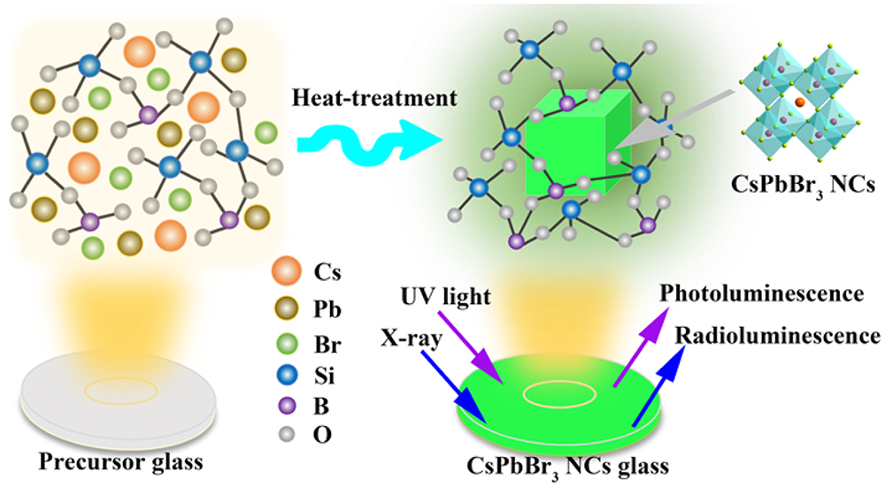
Figure 1.Schematic illustration of the
C. Structural Characterization
The crystal structure of the sample was determined by an X-ray diffractometer (Bruker, D8 Advance) with Cu radiation working at 40 mA and 40 kV. Transmission electron microscopy (TEM, JEM-2100F) was used to investigate the microstructure of NCs. The distribution of elements was analyzed by means of an energy dispersive spectrometer (EDS) attached to TEM. The optical performance of NC glasses was characterized by the combined fluorescence lifetime measurement system (Horiba Jobin Yvon, FluoroMAX-4-TCSPC) and UV-3600 UV-visible spectrometer. The determination of element valence states was collected via an X-ray photoelectron spectrometer (ThermoFisher Scientific ESCALAB250XI). The RL spectra were recorded by a scintillator fluorescence spectrometer (Zolix SBP300) under the excitation source of the X-ray tube of 50 kV. Photos of X-ray imaging were taken by a CCD camera (voltage: 50 kV, dose rate: ).
3. RESULTS AND DISCUSSION
To obtain the optimal optical properties of CPB-0Eu glass, the precursor glasses were heat-treated at 480°C–540°C for 10 h. As shown in Fig. 2(a), the diffraction peak is not obvious and shows only an amorphous halo when the heat-treatment temperature is 480°C for 10 h. With the rise of heat-treatment temperature, X-ray diffraction (XRD) peaks appear by degrees and show a slight redshift, but always correspond well to standard cards, substantiating that the NC phase precipitates successfully in the glass. In addition, the TEM image of CPB-0Eu at 520°C for 10 h shown in Fig. 2(b) shows that the NCs (black spots) are distributed in the glass matrix. Furthermore, a high-resolution (HR) TEM (HRTEM) image in Fig. 2(d) with an unambiguous lattice streak of NCs predicts that the synthesized NCs have excellent crystallinity. It is worth noting that the interplanar spacing of the NCs is 3.05 Å, corresponding to (200) interplanar spacing, and ulteriorly confirms the formation of NCs. The standard structure model diagram of NCs is shown in Fig. 2(e). Figure 2(c) presents the photoluminescence (PL) spectra of glass samples at different crystallization temperatures. Distinctly, the emission peak can be tunable in the range of 523–530 nm, which benefits by the adjustment of heat-treatment temperature. Among them, the CPB-0Eu glass shows the best luminescence intensity at 520°C for 10 h, and its PL quantum yield (PLQY) reaches 56.3%. As shown in the absorption spectra of Fig. 2(f), the position of absorption wavelength shifts to red as the heat-treatment temperature increases, which is consistent with PL spectra. An apparent increment in fluorescence lifetime values with the augmentation of crystallization temperature in Fig. 2(g) is potentially attributed to the observably reduced non-radiative deexcitation effect by the carriers [30].
![]()
Figure 2.(a) XRD patterns of CPB-0Eu at different heat-treatment temperatures. (b) TEM image of CPB-0Eu at 520°C for 10 h. (c) PL spectra under UV light excitation of CPB-0Eu at different heat-treatment temperatures. (d) HRTEM image of CPB-0Eu at 520°C for 10 h. (e) Standard structure model diagram of
Subsequently, a series of -doped glasses (CPB-xEu) were prepared as shown in Fig. 3(a). The four samples under daylight (left) and in UV light (right) provided in Fig. 3(a) can obviously emit high color purity and excellent uniform green light under UV excitation. For the sake of investigating the doping of in CPB-xEu, we made many characterizations of CPB-xEu at 520°C for 10 h. As can be seen in Fig. 3(b), when the concentration of increases, the XRD peaks of the CPB-xEu glasses become more evident and slightly offset toward the larger angle, especially the lattice plane of (110) and (200). It is well known that the Pb-site and X-site in perovskite () determine the size of octahedron. When the Pb ion is replaced by a smaller cation, the size of the octahedron decreases, thereby reducing the size of the A-site cubic octahedron void [31]. Therefore, we hold the opinion that the slight shift of XRD peaks towards higher angles for CPB-xEu glasses is mainly because the larger radius of (119 pm) is partially replaced by the smaller radius of (102.5 pm), resulting in lattice shrinking of NCs [32]. Figures 3(c) and 3(e) present the variation of the bandgap of CPB-xEu glass. In contrast to undoped perovskite glass, the bandgap values increase from 2.362 to 2.400 eV with the gradual increasing of concentration, which further indicates that Eu ions partially enter the perovskite NCs, leading to lattice contraction. In addition, the emission spectra of CPB-xEu glass in Fig. 3(d) also confirm that the fluorescence peak with the assistance of has a blueshift trend, and the emission value of NCs is enhanced with the doping of appropriate content (PLQY of CPB-0.6Eu is 58.6%), which may be attributed to the enhancement of radiative recombination by the defect reduction effect of Eu ions, thereby reducing non-radiative recombination pathways [33]. Ideally, based on the above characterizations, we conclude that part of successfully enters the lattice of NCs.
![]()
Figure 3.(a) Images of CPB-
Similarly, to better verify the microstructure changes of NCs, TEM and XPS (X-ray photoelectron spectroscopy) images are further exhibited in Fig. 4. As shown in Fig. 4(a), NCs are uniformly distributed in the glass matrix, and an HRTEM image of the inset displays a lattice spacing of 2.95 Å, corresponding to the (200) plane. Compared with CPB-0Eu [Fig. 2(d)], the fringe spacing of CPB-1.8Eu on the (200) crystal plane decreases slightly, which is closely related to the entry of into the perovskite lattice, corresponding to XRD results. Moreover, Fig. 4(c) shows the element scan images of the selected glass region, recording the element mapping of Cs, Pb, Br, and Eu. The energy dispersive X-ray (EDX) detector diagrams show that the four (Cs, Pb, Br, and Eu) elements are uniformly distributed in the glass matrix, and verify the existence of in the NCs-at-glasses. To further explore the effect of on NCs-at-glasses, XPS tests were performed on samples CPB-0Eu and CPB-xEu. As shown in Fig. 4(b), the two spectra exhibit the signals of Al 2p, Pb 4f, B 1s, O 1s, Cs 3d, Zn 2p, and Mg 1s derived from the perovskite glass. But unfortunately, it is inconspicuous for the signals of Br 3d and Eu 3d, which may be because they are masked by other stronger signals. Therefore, the corresponding HR spectra of Cs 3d, Pb 4f, Br 3d, Al 2p, and Eu 3d are presented in Figs. 4(d)–4(g) to probe the changes of :Eu NCs-at-glasses more intuitively. It can be seen from the HR-XPS spectra in Figs. 4(d)–4(f), that the binding energy of Cs 3d5/2 and Cs 3d3/2 has no significant variation, including Br 3d5/2 and Br 3d3/2. Also, in the range of 73–80 eV, the spectra of Br 3d3/2 near 75 eV and Al 2p near 75 eV overlap, and their intensities are much greater than that of Br 3d5/2. The HR-XPS spectra of Eu 3d in Fig. 4(g) also verify the presence of Eu ions in the vitreous body. However, the peak of Pb 4f7/2 and Pb 4f5/2 in Fig. 4(e) moves slightly towards the low binding energy, changing from 138.9 to 138.5 eV and 143.5 to 143.3 eV, separately. Hence, we infer that sectional Eu ions incorporate into the lattice [34], which is consistent with the XRD and bandgap results.
![]()
Figure 4.(a) TEM image of CPB-1.8Eu (illustration is the HRTEM image of CPB-1.8Eu at 520°C for 10 h). (b) XPS survey spectra of CPB-0Eu and CPB-1.8Eu. (c) EDX mapping images of CPB-1.8Eu. Corresponding high-resolution spectra of (d) Cs 3d for CPB-0Eu and CPB-1.8Eu, (e) Pb 4f for CPB-
is always identified as an excellent choice to provide red emission for ceramic glass [35]. What is more, -doped luminescent materials (, ) have been applied in Euro ticket checking, which makes it possible to produce visible light under the excitation of a UV lamp [35]. To explore the potential advantages of NCs glass doped with , the samples were further characterized. First, the differences in crystal distribution and particle size based on the incorporation of obtained from TEM images of CPB-0Eu and CPB-1.8Eu in different proportions are presented. As shown in Fig. 5(a), the dark spheres in a glass matrix act as NCs with an average particle size of . In addition, many white spheres, confirmed as pores in previous literature [36], in the TEM image of CPB-0Eu are discovered. They occupy the position of the glass matrix, making the distribution of NCs uneven. Interestingly, the Eu-doped NCs shrink significantly, and the average diameter decreases to , along with a more uniform distribution without pores [Fig. 5(b)]. Therefore, we think that the added Eu ions could be identified as a nucleating agent to promote the nucleation of NCs, thus reducing the crystal size and improving the distribution uniformity of NCs in glass [37]. Worthy of note, when designing the ratio of glass components, it plays a decisive role for the concentration of perovskite to control the optical property and transparency of perovskite glass, which are important indicators to ensure effective X-ray absorption. This is because poor transparency can cause light scattering to seriously affect the imaging resolution. Herein, the thickness of all test samples was , and we monitored the transmittance spectra of them to assess their light scattering phenomenon [Fig. 5(c)]. The comparison of the undoped- and doped- glasses shows that the former is dominated by scattering, while the latter is dominated by absorption, indicating that the addition of Eu ions can significantly reduce the light scattering of pure perovskite glass. It also provides a foundation for obtaining an HR image. In addition, Fig. 5(e) visually shows that the transparency of CPB-xEu gradually increases with the embedding of an appropriate amount of , in agreement with the above results. PL intensities were measured at different locations of the same sample to evaluate uniformity of samples. We took the value measured at the first position as the standard, and plotted a linear plot as shown in Fig. 8 (Appendix A). Compared with the undoped glass whose PL relative difference is , the -doped NC glass shows a smaller relative difference of PL values, which have almost no change, similar to the results of the particle size distribution obtained by TEM. This also predicts that the -doped NCs glasses have enhanced uniformity. Furthermore, in contrast to other literatures on Eu-doped glass [34,38–40], the PL signals of under UV light excitation based on -doped glass prepared in this work are almost inhibited, so we recorded the excitation spectra of CPB-xEu glasses to explore the most effective excitation wavelength for CPB-xEu in Fig. 9(a) (Appendix A). All the samples display a wide peak near 360 nm, which is considered to be the optimal excitation wavelength for NCs. What is more, the PL spectra of CPB-xEu glasses excited at 355–395 nm are collected as shown in Figs. 9(b)–9(e) (Appendix A). Obviously, with the increase in excitation wavelength (energy decrease), the inherent emission intensity of NCs at about 525 nm gradually decreases. In consideration of the emission and taking CPB-0.6Eu for example, the partial enlargement spectra of emission based on Fig. 9(c) (Appendix A) under 355–395 nm excitation are given in Fig. 10 (Appendix A), which exhibit significant Eu ion emission (613 nm, ) except for 355 nm under excitation of 355–395 nm. Therefore, due to the above phenomenon, we chose 365 nm as the most appropriate excitation wavelength to excite -doped glasses. Interestingly, CPB-xEu glasses have no characteristic peak of at various wavelengths of excitation [Figs. 9(b)–9(e) in Appendix A]. We reason that the amount of is very small relative to NCs (25:0.4–1.8), so NCs are the major contributors of PL spectra. Figure 5(d) displays the characteristic emission of Eu ions in the range of 590–700 nm (a partial enlargement of the illustration), and the signal of is particularly strong at 613 nm and 654 nm, which can be assigned to and , correspondingly. To be precise, if the emission peaks of NCs are filtered, the characteristic peaks of Eu ions can also be observed. Figure 11 (Appendix A) provides the decay life of CPB-0Eu and CPB-0.6Eu, which is 43.95 ns and 49.17 ns, respectively, and still in nanosecond scope.
![]()
Figure 5.TEM images and histograms of size distribution of (a) CPB-0Eu and (b) CPB-1.8Eu. (c) Transmittance spectra of CPB-
To evaluate the potential scintillation properties of CPB-xEu, the RL spectra of CPB-xEu (, 0.6, 1.8) glass were investigated at a radiation dose of and voltage of 50 kV in Figs. 6(a)–6(c). Evidently, RL spectra show that the emission values of the samples gradually increase, when the voltage remains constant (50 kV) and the current increases from 10 to 30 μA. In addition, it is pleasantly surprising for us that the intensity of the emission peak has no significant saturation in the range where the tube current increases from 10 to 30 μA, which indicates that the CPB-xEu have a strong sensitivity to X-ray detection. By comparing Figs. 6(a) and 6(b), we found that the emission value doubles when 0.6% Eu is incorporated into perovskite glass, predicting that a certain addition of rare earth could enhance the RL property of glass, which is similar to the PL results. In detail, the intrinsic peaks of fluorescence emission of NCs excited by X-rays of different tube currents are all located at about 528 nm in Figs. 6(a)–6(c). Interestingly, CPB-0.6Eu and CPB-1.8Eu have multimodal luminescence, which is attributed to the typical f-f transitions of ions. As illustrated in Figs. 6(b) and 6(c), five distinct emission peaks of ions are situated at 577 nm (), 590 nm (), 612 nm (), 651 nm (), and 700 nm () [32]. When the concentration of Eu is 1.8, the emission of is dominant, especially for the radiative transition of . This unique property of transitions of ions gives perovskite with a direct bandgap the advantage of reducing the overlap between RL spectra and absorption, resulting in a decrease in the probability of self-absorption (Fig. 12 in Appendix A). Furthermore, according to the RL integral intensity of CPB-xEu (, 0.6, 1.8) at the same voltage and different radiation dose rates (different tube currents), we have plotted the function relationship between the RL integral intensity of CPB-xEu (, 0.6, 1.8) and the dose rate of the incident X-ray. As shown in Fig. 13 (Appendix A), the functions of the three samples are superlinear, which is conducive to acquiring outstanding X-ray images. The superlinear relationship (degree of fitting is the same) among the three samples was almost alike, which may be because the radiation dose that the samples can endure has not reached the saturation state.
![]()
Figure 6.RL spectra of (a) CPB-0Eu, (b) CPB-0.6Eu, and (c) CPB-1.8Eu upon increasing current of X-ray cathode tube. (d), (e) Schematic illustration of RL mechanism about CPB-
Based on the unique scintillation property of :Eu NCs-at-glasses, we propose their mechanism of X-ray scintillation [see Figs. 6(d) and 6(e)]. Generally, in an emblematic scintillator material, high-energy photons generated by X-rays can interact with heavier atoms (such as Pb, Tl, Ce, or Eu) through the photoelectric effect to produce a considerable amount of hot electrons [41]. Similarly, in the NCs-at-glasses, X-ray photons mainly interact with the lattice atoms of the NCs. During the process, abundant electrons and holes are induced, and electron transport occurs in the NCs. In this case, these electrons (yellow circles, ) will be ejected from the lattice atoms, resulting in high-energy secondary electrons. The procreant thermal carriers (electrons and holes) undergo subsequent thermalization to generate low-energy excitons. Then, the ideal visible RL is produced by rapid radiative recombination of the thermalized electron/hole to the conduction/valence band edge [22,42]. For some of the Eu ions that do not occupy a Pb ion position, it is an independent RL center for -doped glass. After ionizing radiation, the generated electrons and holes are transmitted in their valence band and conduction band, respectively. Subsequently, directly captures carriers and recombines with holes or radiation to form excitons, and then produces a transition to emit light [43].
Subsequently, we compared and investigated the change of integrated radiation intensity dependence on the time of CPB-0.6Eu and CPB-0Eu glasses at a radiation dose of and a voltage of 50 kV. Surprisingly, the RL values for CPB-0.6Eu remain at almost 100% of the original RL intensity following radiation exposure for 25 h. But for CPB-0Eu, it retains 84% of its original value after 25 h of radiation. In addition, we compared the dissolution of the samples immersed in water for five days before and after radiation for 25 h (Fig. 14 in Appendix A). It is obvious that although CPB-0.6Eu was exposed to X-rays for up to 25 h and then soaked in water for five days, the amount of Pb leaching from 10.56 to 10.74 μg/L remained relatively stable. At the same time, CPB-0Eu without Eu showed relatively weaker stability whose dissolution increased from 12.32 to 13.46 μg/L. This further indicates that the robust glass acts as the inner protective matrix of NCs, which endows the NCs glass with super stability. More importantly, we can prove that Eu-doped NCs have more excellent stability because the lattice parameters of NCs slightly reduce and cohesion increases [44].
Benefiting from the optimized luminescence properties, enhanced uniformity, and attenuated scattering of the CPB-xEu (, 0.6, 1.8) glasses prepared, we construct a simple imaging system to investigate the application performance of a perovskite scintillator in X-ray imaging [Fig. 7(a)]. Primarily, the lightproof samples containing a spring are placed between the X-ray source and a platform with a perovskite glass scintillator [see Figs. 7(b) and 7(c) for details of the opaque sample], and X-rays are directed at the opaque side (side view) of the object. Finally, a camera is installed to capture X-ray images. As shown in Figs. 7(d)–7(f), the X-ray images ideally display the basic structure of the inner spring, so it can indicate that the perovskite glass X-ray imaging system is capable of detecting the internal structure of the object. Interestingly, with the increase in concentration, the images excited by X-rays turn to red by degrees, which is caused by the superposition of green emitting and red emitting : . The X-ray images became clearer due to the weakening of light scattering. For a pure NC glass scintillator with poor RL emission and transparency, the obtained image was not particularly ideal. On this basis, we found that the addition of to NC glass is indeed an effective strategy for enhancing the RL emission of a scintillator, reducing light scattering, along with increasing its transparency. CPB-0.6Eu achieved strong multi-mode luminescence of 500–725 nm while ensuring outstanding luminescence of NCs, and also obtained legible X-ray images. For CPB-1.8Eu, although the X-ray image was the most obvious, the emission of NCs was almost covered by the intense Eu ion luminescence due to the increase in the content of Eu, which was dominated by the transition of . This could not be attributed to NCs. Therefore, we concluded that in the -doped glass scintillator system, in addition to luminescence of , the intrinsic luminescence of NCs was very crucial, which determined its RL characteristics. As such, -doped glass is promising to be an underlying candidate for X-ray detection and imaging.
![]()
Figure 7.(a) Schematic of the experimental setup used for X-ray imaging with sample (X-rays are excited directly at the side of the sample; voltage: 50 kV, dose rate:
4. CONCLUSION
In summary, a series of enhanced multimodal luminescence CPB-xEu glasses with prominent PLQY and good uniformity was acquired by conventional high-temperature sintering and subsequent heat treatment. Surprisingly, the emission peak of the fabricated CPB-xEu glasses appeared close to 528 nm and maintained excellent luminescence. We confirmed the successful incorporation of into the NCs lattice by various structural characterizations. In addition, the advantages of doping Eu in NCs glass have been evaluated. Owing to enhanced uniformity, transparency, along with reduced scattering, the addition of effectively enhances the RL intensity. Finally, the constructed X-ray imaging system demonstrated that the -doped perovskite glass scintillator has far-reaching application values in X-ray imaging and detection. In particular, multimodal luminescence of glasses also furnishes a new thought for X-ray anti-counterfeiting, opening the door to amplify the application of glass.
APPENDIX A
Figure
![]()
Figure 8.Relative PL intensity at five different locations of the same sample.
![]()
Figure 9.(a) Excitation spectra of CPB-
![]()
Figure 10.Characteristic PL spectra of Eu ions at 560–700 nm under different wavelength excitation of CPB-0.6Eu.
![]()
Figure 11.PL decay of CPB-0Eu and CPB-0.6Eu glasses.
![]()
Figure 12.RL spectra of CPB-
![]()
Figure 13.Linear relation based on RL intensity and X-ray dose rate.
![]()
Figure 14.The relative RL intensity of CPB-0.6Eu and CPB-0Eu glass depending on time of X-ray radiation at a radiation dose of 105 μGyair/s and a voltage of 50 kV. The illustration shows a histogram of Pb2+ solubility after 5 days of water immersion before and after X-ray radiation.
References
[1] A. Horimoto, N. Kawano, D. Nakauchi, H. Kimura, M. Akatsuka, T. Yanagida. Scintillation properties of organic–inorganic perovskite-type compounds with fluorophenethylamine. Opt. Mater., 101, 109686(2020).
[2] D. Yu, P. Wang, F. Cao, Y. Gu, J. Liu, Z. Han, B. Huang, Y. Zou, X. Xu, H. Zeng. Two-dimensional halide perovskite as β-rayscintillator for nuclear radiation monitoring. Nat. Commun., 11, 3395(2020).
[3] F. Maddalena, L. Tjahjana, A. Xie, , S. Zeng, H. Wang, P. Coquet, W. Drozdowski, C. Dujardin, C. Dang, M. Birowosuto. Inorganic, organic, and perovskite halides with nanotechnology for high–light yield X- and γ-ray scintillators. Crystals, 9, 88(2019).
[4] K. S. Kim, S. Y. Kang, W. S. Kim, H. S. Cho, C. K. Park, D. Y. Lee, G. A. Kim, S. Y. Park, H. W. Lim, H. W. Lee, J. E. Park, D. H. Jeon, Y. H. Lim, U. K. Je, T. H. Woo. Improvement of radiographic visibility using an image restoration method based on a simple radiographic scattering model for X-ray nondestructive testing. NDT & E Int., 98, 117-122(2018).
[5] P. Lecoq. Development of new scintillators for medical applications. Nucl. Instrum. Methods Phys. Res. A, 809, 130-139(2016).
[6] H. Wei, J. Huang. Halide lead perovskites for ionizing radiation detection. Nat. Commun., 10, 1066(2019).
[7] A. Phunpueok, V. Thongpool, S. Jaiyen. Scintillation response of CdWO4 scintillator for gamma-ray detection. J. Phys. Conf. Ser., 1380, 012128(2019).
[8] M. Tyagi, A. Khan, P. Q. Vuong, H. J. Kim. Comprehending the role of trap centers and host energy transfers in excitation density dependent kinetics of Ce doped Gd3Ga3Al2O12 scintillator; an unresolved scintillation characteristic. J. Lumin., 219, 116815(2020).
[9] Y. Wu, J. Peng, D. Rutstrom, M. Koschan, C. Foster, C. L. Melcher. Unraveling the critical role of site occupancy of lithium codopants in Lu2SiO5:Ce3+ single-crystalline scintillators. ACS. Appl. Mater. Interfaces, 11, 8194-8201(2019).
[10] A. Khan, D. J. Daniel, M. Tyagi, H. J. Kim, M. H. Lee, Y. Kim. Czochralski growth, electronic structure, luminescence and scintillation properties of Cs2Mo3O10: a new scintillation crystal for ovββ decay search. J. Alloys Comp., 821, 153466(2020).
[11] K. Kagami, M. Koshimizu, Y. Fujimoto, S. Kishimoto, R. Haruki, F. Nishikido, K. Asai. High-energy X-ray detection capabilities of Hf-loaded plastic scintillators synthesized by sol–gel method. J. Mater. Sci., 31, 896-902(2019).
[12] B. K. Cha, J. Y. Kim, T. J. Kim, C. Sim, G. Cho. Fabrication and imaging characterization of high sensitive CsI(Tl) and Gd2O2S(Tb) scintillator screens for X-ray imaging detectors. Radiat. Meas., 45, 742-745(2010).
[13] O. Guillot-Noël, J. T. M. de Haas, P. Dorenbos, C. W. E. van Eijk, K. Krämer, H. U. Güdel. Optical and scintillation properties of cerium-doped LaCl3, LuBr3 and LuCl3. J. Lumin., 85, 21-35(1999).
[14] A. S. Pushak, V. V. Vistovskyy, T. M. Demkiv, A. A. Halyatkin, Y. R. Datsyuk, A. V. Gektin, S. V. Myagkota, P. A. Rodnyi, A. S. Voloshinovskii. Luminescence of SrX2 microcrystals (X = Cl, I) activated with Eu2+ ions and dispersed in a NaI matrix. Opt. Spectrosc., 117, 593-597(2014).
[15] J. M. Ogieglo, A. Katelnikovas, A. Zych, T. Justel, A. Meijerink, C. R. Ronda. Luminescence and luminescence quenching in Gd3(Ga, Al)5O12 scintillators doped with Ce3+. J. Phys. Chem. A., 117, 2479-2484(2013).
[16] M. Mori, J. Xu, G. Okada, T. Yanagida, J. Ueda, S. Tanabe. Comparative study of optical and scintillation properties of Ce:YAGG, Ce:GAGG and Ce:LuAGG transparent ceramics. J. Ceram. Soc. Jpn., 124, 569-573(2016).
[17] A. Xie, T. H. Nguyen, C. Hettiarachchi, M. E. Witkowski, W. Drozdowski, M. D. Birowosuto, H. Wang, C. Dang. Thermal quenching and dose studies of X-ray luminescence in single crystals of halide perovskites. J. Phys. Chem. C, 122, 16265(2018).
[18] C. Ji, S. Wang, Y. Wang, H. Chen, L. Li, Z. Sun, Y. Sui, S. Wang, J. Luo. 2D hybrid perovskite ferroelectric enables highly sensitive X-ray detection with low driving voltage. Adv. Funct. Mater., 30, 1905529(2019).
[19] W. Pan, B. Yang, G. Niu, K. H. Xue, X. Du, L. Yin, M. Zhang, H. Wu, X. S. Miao, J. Tang. Hot-pressed CsPbBr3 quasi-monocrystalline film for sensitive direct X-ray detection. Adv. Mater., 31, 1904405(2019).
[20] F. R. Hu, H. C. Zhang, C. Sun, C. Y. Yin, B. H. Lv, C. F. Zhang, W. W. Yu, X. Y. Wang, Y. Zhang, M. Xiao. Superior optical properties of perovskite nanocrystals as single photon emitters. ACS. Nano., 9, 12410-12416(2015).
[21] J. H. Heo, D. H. Shin, J. K. Park, D. H. Kim, S. J. Lee, S. H. Im. High-performance next-generation perovskite nanocrystal scintillator for nondestructive X-ray imaging. Adv. Mater., 30, 1801743(2018).
[22] Q. Chen, J. Wu, X. Ou, B. Huang, J. Almutlaq, A. A. Zhumekenov, X. Guan, S. Han, L. Liang, Z. Yi, J. Li, X. Xie, Y. Wang, Y. Li, D. Fan, D. B. L. Teh, A. H. All, O. F. Mohammed, O. M. Bakr, T. Wu, M. Bettinelli, H. Yang, W. Huang, X. Liu. All-inorganic perovskite nanocrystal scintillators. Nature, 561, 88-93(2018).
[23] C. C. Stoumpos, C. D. Malliakas, J. A. Peters, Z. Liu, M. Sebastian, J. Im, T. C. Chasapis, A. C. Wibowo, D. Y. Chung, A. J. Freeman, B. W. Wessels, M. G. Kanatzidis. Crystal growth of the perovskite semiconductor CsPbBr3: a new material for high-energy radiation detection. Cryst. Growth Des., 13, 2722-2727(2013).
[24] C. Zheng, C. Bi, F. Huang, D. Binks, J. Tian. Stable and strong emission CsPbBr3 quantum dots by surface engineering for high-performance optoelectronic films. ACS Appl. Mater. Interfaces, 11, 25410-25416(2019).
[25] F. Cao, D. Yu, W. Ma, X. Xu, B. Cai, Y. M. Yang, S. Liu, L. He, Y. Ke, S. Lan, K. L. Choy, H. Zeng. Shining emitter in a stable host: design of halide perovskite scintillators for X-ray imaging from commercial concept. ACS Nano, 14, 5183-5193(2020).
[26] C. Wang, H. Lin, Z. Zhang, Z. Qiu, H. Yang, Y. Cheng, J. Xu, X. Xiang, L. Zhang, Y. Wang. X-ray excited CsPb(Cl,Br)3 perovskite quantum dots-glass composite with long-lifetime. J. Eur. Ceram. Soc., 40, 2234-2238(2020).
[27] B. Zhang, K. Zhang, L. Li, C. Xu, R. Wang, C. Wang, J. Yang, Y. Yang, J. Wang, F. Qiu, T. Sun, C. Zhou, X. Wen. Enhancing stability and luminescence quantum yield of CsPbBr3 quantum dots by embedded in borosilicate glass. J. Alloy. Compd., 874, 159962(2021).
[28] P. Aryal, H. Kima, S. Saha, J. Cho, A. V. Ntarisa, S. Kothan, J. Kaewkhao. Rapid and convenient crystallization of quantum dot CsPbBr3 inside a phosphate glass matrix. J. Alloy. Compd., 866, 158974(2021).
[29] Y. Lu, P. Li, W. Xie, Y. Duan, X. Wang, S. Xu, J. Zhang. Negative thermal quenching CsPbBr3 glass-ceramic based on intrinsic radiation and vacancy defect co-induced dual-emission. J. Eur. Ceram. Soc., 41, 3635-3642(2021).
[30] Y. Ye, W. Zhang, Z. Zhao, J. Wang, C. Liu, Z. Deng, X. Zhao, J. Han. Highly luminescent cesium lead halide perovskite nanocrystals stabilized in glasses for light-emitting applications. Adv. Opt. Mater., 7, 1801663(2019).
[31] C. Shen, Y. Zhao, L. Yuan, L. Ding, Y. Chen, H. Yang, S. Liu, J. Nie, W. Xiang, X. Liang. Transition metal ion doping perovskite nanocrystals for high luminescence quantum yield. Chem. Eng. J., 382, 122868(2020).
[32] C. Daqin, F. Gaoliang, C. Xiao, L. Lei, Z. Jiasong, M. Qinan, Z. Su, L. Junni. Mn Doped CsPbCl3 perovskite nanocrystals: solvothermal synthesis, dual-color luminescence and improved stability. J. Phys. Chem. C, 6, 8990-8998(2018).
[33] C. M. Guvenc, Y. Yalcinkaya, S. Ozen, H. Sahin, M. M. Demir. Gd3+-doped α-CsPbI3 nanocrystals with better phase stability and optical properties. J. Phys. Chem. C, 123, 24865-24872(2019).
[34] P. Li, Y. Duan, Y. Lu, A. Xiao, Z. Zeng, S. Xu, J. Zhang. Nanocrystalline structure control and tunable luminescence mechanism of Eu-doped CsPbBr3 quantum dot glass for WLEDs. Nanoscale, 12, 6630-6636(2020).
[35] C. Wu, S. Zeng, Z. Wang, F. Wang, H. Zhou, J. Zhang, Z. Ci, L. Sun. Efficient mechanoluminescent elastomers for dual-responsive anticounterfeiting device and stretching/strain sensor with multimode sensibility. Adv. Funct. Mater., 28, 1803168(2018).
[36] P. Li, Y. Lu, Y. Duan, S. Xu, J. Zhang. Potential application of perovskite glass material in photocatalysis field. J. Phys. Chem. C, 125, 2382-2392(2021).
[37] W. Ma, T. Jiang, Z. Yang, H. Zhang, Y. Su, Z. Chen, X. Chen, Y. Ma, W. Zhu, X. Yu, H. Zhu, J. Qiu, X. Liu, X. Xu, Y. Yang. Highly resolved and robust dynamic X-ray imaging using perovskite glass-ceramic scintillator with reduced light scattering. Adv. Sci., 8, 2003728(2021).
[38] G. Pan, X. Bai, D. Yang, X. Chen, P. Jing, S. Qu, L. Zhang, D. Zhou, J. Zhu, W. Xu, B. Dong, H. Song. Doping lanthanide into perovskite nanocrystals: highly improved and expanded optical properties. Nano Lett., 17, 8005-8011(2017).
[39] R. Yuan, L. Shen, C. Shen, J. Liu, L. Zhou, W. Xiang, X. Liang. CsPbBr3:
[40] Y. Cheng, C. Shen, L. Shen, W. Xiang, X. Liang. Tb3+, Eu3+ co-doped CsPbBr3 QDs glass with highly stable and luminous adjustable for white LEDs. ACS Appl. Mater. Interfaces, 10, 21434-21444(2018).
[41] M. J. Weber. Inorganic scintillators: today and tomorrow. J. Lumin., 100, 35-45(2002).
[42] F. Zhou, Z. Li, W. Lan, Q. Wang, L. Ding, Z. Jin. Halide perovskite, a potential scintillator for X-ray detection. Small Methods, 4, 2000506(2020).
[43] F. Moretti, G. Patton, A. Belsky, M. Fasoli, A. Vedda, M. Trevisani, M. Bettinelli, C. Dujardin. Radioluminescence sensitization in scintillators and phosphors: trap engineering and modeling. J. Phys. Chem. C, 118, 9670-9676(2014).
[44] G. E. Eperon, D. S. Ginger. B-site metal cation exchange in halide perovskites. ACS Energy Lett., 2, 1190-1196(2017).

Set citation alerts for the article
Please enter your email address
© Copyright 2018-2021 | Chinese Laser Press. All Rights Reserved 沪ICP备15018463号-20