• Spectroscopy and Spectral Analysis
  • Vol. 42, Issue 9, 2983 (2022)
Nan SUN, Hong-lin TAN*;, Zheng-dong ZHANG, Xiang REN, Yan ZHOU, Jian-qi LIU, Xiao-ming CAI, and Jin-ming CAI
Author Affiliations
  • Faculty of Materials Science and Engineering, Kunming University of Science and Technology, Kunming 650093, China
  • show less
    DOI: 10.3964/j.issn.1000-0593(2022)09-2983-06 Cite this Article
    Nan SUN, Hong-lin TAN, Zheng-dong ZHANG, Xiang REN, Yan ZHOU, Jian-qi LIU, Xiao-ming CAI, Jin-ming CAI. Raman Spectroscopy Analysis and Formation Mechanism of Carbon Nanotubes Doped Polyacrylonitrile/Copper Cyclized to Graphite at Room Temperature[J]. Spectroscopy and Spectral Analysis, 2022, 42(9): 2983 Copy Citation Text show less
    (a) Raman spectra of PAN powder and 0.4% PAN-DMF solution; (b) Raman spectra of PAN obtained by smooth copper and copper sheet with nitric acid etching 5~25 min annealing at 40 ℃ for 6 h; (c) SEM images of copper substrate after etching with nitric acid for 15 minutes
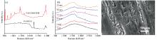
    Fig. 1. (a) Raman spectra of PAN powder and 0.4% PAN-DMF solution; (b) Raman spectra of PAN obtained by smooth copper and copper sheet with nitric acid etching 5~25 min annealing at 40 ℃ for 6 h; (c) SEM images of copper substrate after etching with nitric acid for 15 minutes
    (a, b) Raman and Infrared spectra obtained by annealing at different temperatures of 40~160 ℃ for 6 h;(c) Raman spectra obtained by annealing at 140 ℃ for 1~12 h
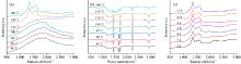
    Fig. 2. (a, b) Raman and Infrared spectra obtained by annealing at different temperatures of 40~160 ℃ for 6 h;(c) Raman spectra obtained by annealing at 140 ℃ for 1~12 h
    (a) Raman spectrum of CNT (black) and nitric acid modified CNT (red); (b) SEM image of PAN-CNT; (c) Raman spectrum of PAN-CNT on SiO2; (d) Raman spectra of PAN-CNT on rough copper sheet irradiated with different power lasers; (e) Raman plan view of PAN-CNT under laser irradiation; (f) SEM image corresponding to the PAN-CNT laser irradiation area in contact with the rough copper
    Fig. 3. (a) Raman spectrum of CNT (black) and nitric acid modified CNT (red); (b) SEM image of PAN-CNT; (c) Raman spectrum of PAN-CNT on SiO2; (d) Raman spectra of PAN-CNT on rough copper sheet irradiated with different power lasers; (e) Raman plan view of PAN-CNT under laser irradiation; (f) SEM image corresponding to the PAN-CNT laser irradiation area in contact with the rough copper
    (a) Raman spectra of PAN-CNT under different conditions; (b) Raman spectra of PAN under different conditions; (c) Schematic diagram of PAN graphitization mechanism on copper sheet; (d) The charge transfer mechanism of CNT doping; (e) Schematic diagram of PAN graphitization driven by thermal electrons
    Fig. 4. (a) Raman spectra of PAN-CNT under different conditions; (b) Raman spectra of PAN under different conditions; (c) Schematic diagram of PAN graphitization mechanism on copper sheet; (d) The charge transfer mechanism of CNT doping; (e) Schematic diagram of PAN graphitization driven by thermal electrons
    Nan SUN, Hong-lin TAN, Zheng-dong ZHANG, Xiang REN, Yan ZHOU, Jian-qi LIU, Xiao-ming CAI, Jin-ming CAI. Raman Spectroscopy Analysis and Formation Mechanism of Carbon Nanotubes Doped Polyacrylonitrile/Copper Cyclized to Graphite at Room Temperature[J]. Spectroscopy and Spectral Analysis, 2022, 42(9): 2983
    Download Citation