• Acta Photonica Sinica
  • Vol. 49, Issue 8, 0817002 (2020)
Le PENG1, Lu ZHOU2, Yan-ping QING3, Li-ying TONG1, Zhao-heng LIANG1, Hao FU1, and Jun ZHOU1、*
Author Affiliations
  • 1Department of Microelectronics Engineering, School of Physical & Science, Ningbo University, Ningbo, Zhejiang 315211, China
  • 2Center for Terahertz Waves, College of Precision Instrument and Optoelectronics Engineering, Tianjin University, Tianjin 300072, China
  • 3Affiliated Hospital, School of Medicine, Ningbo University, Ningbo, Zhejiang 315020, China
  • show less
    DOI: 10.3788/gzxb20204908.0817002 Cite this Article
    Le PENG, Lu ZHOU, Yan-ping QING, Li-ying TONG, Zhao-heng LIANG, Hao FU, Jun ZHOU. High Sensitivity Detection of Gene-like Tumor Markers Based on SERS Characteristics of Hollow Sea-urchin Gold Nanoparticles and Noble Metal/semiconductor Substrate[J]. Acta Photonica Sinica, 2020, 49(8): 0817002 Copy Citation Text show less
    Construction of the sandwich structure detect platform and SERS-based bioassay protocol
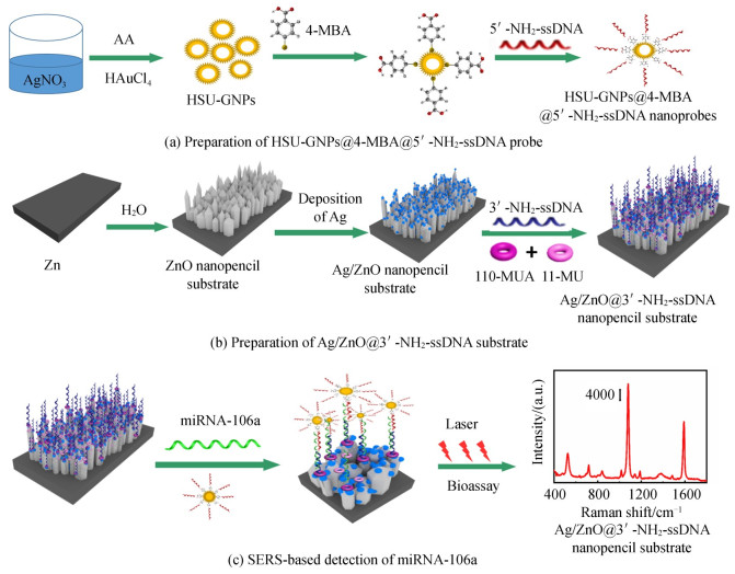
    Fig. 1. Construction of the sandwich structure detect platform and SERS-based bioassay protocol
    SEM and TEM images, EDS spectrum of HSU-GNPs, and the UV-vis absorption spectra of HSU-GNPs, HSU-GNPs@4-MBA and HSU-GNPs@4-MBA@5'-NH2-ssDNA probes.
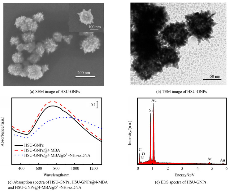
    Fig. 2. SEM and TEM images, EDS spectrum of HSU-GNPs, and the UV-vis absorption spectra of HSU-GNPs, HSU-GNPs@4-MBA and HSU-GNPs@4-MBA@5'-NH2-ssDNA probes.
    SEM images of the ZnO nano-pencil structure and Ag/ZnO nanostructure prepared at different magnetron sputtering time
    Fig. 3. SEM images of the ZnO nano-pencil structure and Ag/ZnO nanostructure prepared at different magnetron sputtering time
    X-rays diffraction patterns of ZnO and Ag/ZnO substrates
    Fig. 4. X-rays diffraction patterns of ZnO and Ag/ZnO substrates
    SERS spectra and the intensities of SERS peaks of 4-MBA attached on various Ag/ZnO nanostructures, ) Raman spectrum of 4-MBA powder (line a), SERS spectra of 4-MBA on the freshly prepared and saved Ag/ZnO (line b and c)
    Fig. 5. SERS spectra and the intensities of SERS peaks of 4-MBA attached on various Ag/ZnO nanostructures, ) Raman spectrum of 4-MBA powder (line a), SERS spectra of 4-MBA on the freshly prepared and saved Ag/ZnO (line b and c)
    SERS spectra of 4-MBA and the dose-response curves corresponding for the miRNA-106a samples
    Fig. 6. SERS spectra of 4-MBA and the dose-response curves corresponding for the miRNA-106a samples
    SERS spectra corresponding to different analytes
    Fig. 7. SERS spectra corresponding to different analytes
    NameSequences
    miRNA-106a5'-AAAAGUGCUUACAGUGCAGGUAG-3'
    Non-complementary-miRNA-215'-UAGCUUAUCAGACUGAUGUUGA-3'
    Non-complementary-miRNA mimics5'-UUGUACUACACAAAAGUACUG-3'
    5'-NH2-ssDNA5'-NH2-AAAAACTACCTICACT-3'
    3'-NH2-ssDNA5'-CTAAICACTTTTAAAAA-NH2-3'
    Table 1. Oligonucleotides sequences
    SampleSERSRT-qPCRRelative deviation
    17.651 pmol·L-17.493 pmol·L-12.06%
    235.829 fmol·L-134.815 fmol·L-12.83%
    322.702 fmol·L-125.353 fmol·L-1-11.67%
    415.635 fmol·L-115.541 fmol·L-10.06%
    513.724 fmol·L-114.216 6 fmol·L-1-3.59%
    62.058 2 fmol·L-1--
    Table 2. Detection results of miRNA-106a
    Le PENG, Lu ZHOU, Yan-ping QING, Li-ying TONG, Zhao-heng LIANG, Hao FU, Jun ZHOU. High Sensitivity Detection of Gene-like Tumor Markers Based on SERS Characteristics of Hollow Sea-urchin Gold Nanoparticles and Noble Metal/semiconductor Substrate[J]. Acta Photonica Sinica, 2020, 49(8): 0817002
    Download Citation