• Laser & Optoelectronics Progress
  • Vol. 55, Issue 4, 041102 (2018)
Tao Wang1、2、*, Minghao Zhang1, Ling Guo1, Wenliang Li1, Jianquan Yao1, Yanliang Zhao1, Xingguo Cao1, Jun Cai1, and Yuxiang Li1
Author Affiliations
  • 1 College of Mechanical Engineering, Hebei University of Technology, Tianjin 300130, China
  • 1 Department of Brain Oncology, Tianjin Medical University Cancer Institute and Hospital, Tianjin 300060, China
  • 1 Hospital of Hebei University of Technology, Tianjin 300130, China
  • 1 Institute of Laser & Opto-Electronics, School of Precision Instrument and Opto-Electronics Engineering, Tianjin University, Tianjin 300130, China;
  • 1 School of Mechanical Engineering, Tianjin Polytechnic University, Tianjin 300387, China
  • 2 Institute of Laser & Opto-Electronics, School of Precision Instrument and Opto-Electronics Engineering, Tianjin University, Tianjin 300130, China;
  • show less
    DOI: 10.3788/LOP55.041102 Cite this Article Set citation alerts
    Tao Wang, Minghao Zhang, Ling Guo, Wenliang Li, Jianquan Yao, Yanliang Zhao, Xingguo Cao, Jun Cai, Yuxiang Li. Research on Dual-Mode Biomedical Imaging Based on Free-Form Surface Optics[J]. Laser & Optoelectronics Progress, 2018, 55(4): 041102 Copy Citation Text show less
    Relationship between focal power of tumor lesion tissue and viewpoint spacing
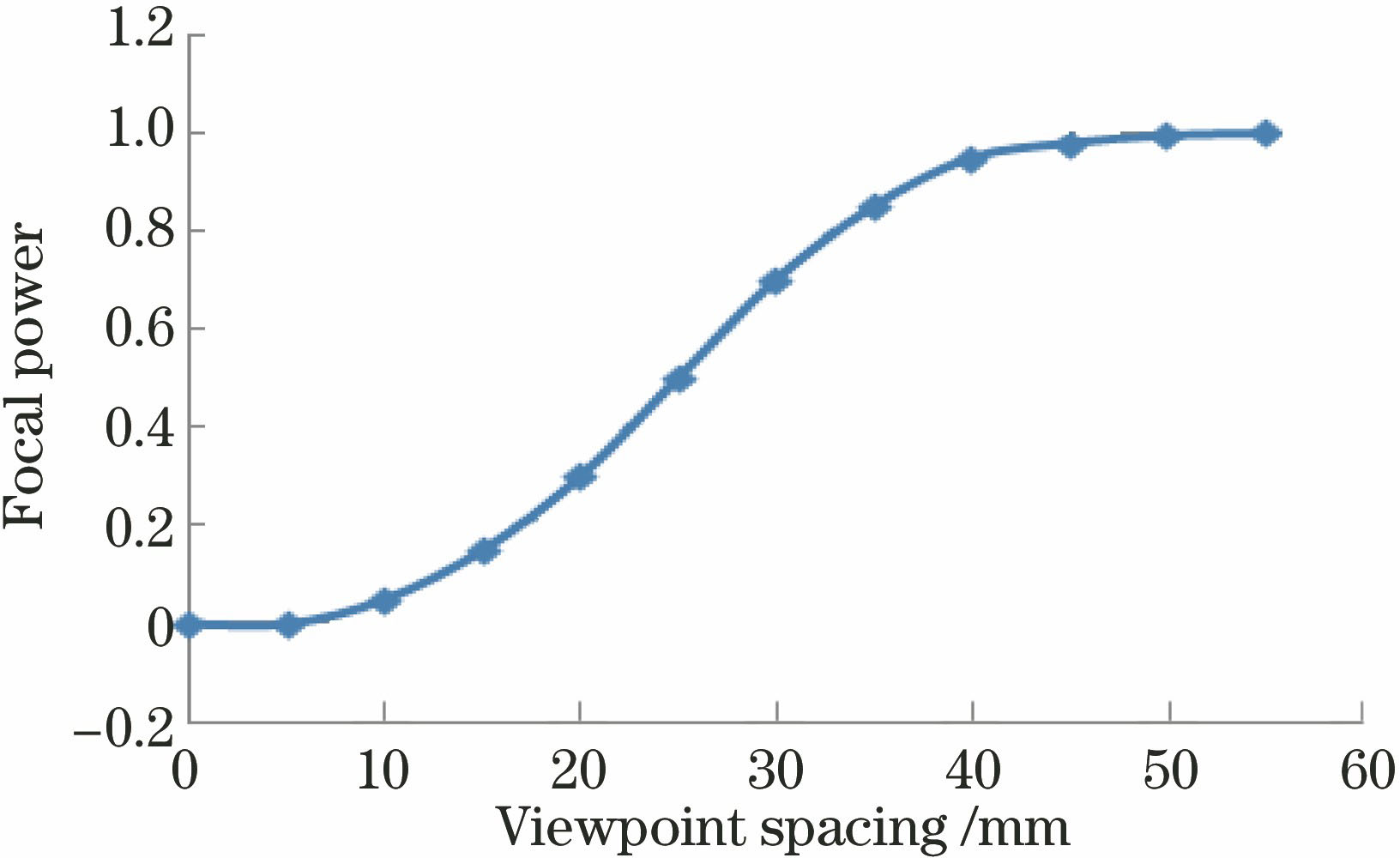
    Fig. 1. Relationship between focal power of tumor lesion tissue and viewpoint spacing
    Numerical simulation of free-form surface optical characteristic channel of lesion tissue
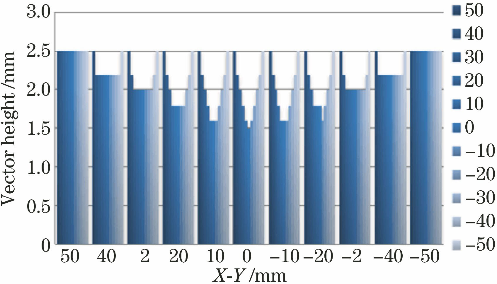
    Fig. 2. Numerical simulation of free-form surface optical characteristic channel of lesion tissue
    Three-dimensional numerical simulation of free-form surface optical channel lens and imaging surface of lesion tissue
    Fig. 3. Three-dimensional numerical simulation of free-form surface optical channel lens and imaging surface of lesion tissue
    Free-form surface optical diagram of optical molecular image and CT image
    Fig. 4. Free-form surface optical diagram of optical molecular image and CT image
    Processed three-dimensional data envelope of free-form surface lens and imaging surface of lesion tissue
    Fig. 5. Processed three-dimensional data envelope of free-form surface lens and imaging surface of lesion tissue
    Mapping of free-form surface optical characteristic channel of lesion tissue
    Fig. 6. Mapping of free-form surface optical characteristic channel of lesion tissue
    Free-form surface optical coupling image of lesion tissue
    Fig. 7. Free-form surface optical coupling image of lesion tissue
    Signal-to-noise ratio of free-form surface optical image signal of lesion tissue
    Fig. 8. Signal-to-noise ratio of free-form surface optical image signal of lesion tissue
    Common image of lesion tissue
    Fig. 9. Common image of lesion tissue
    Signal-to-noise ratio of common image of lesion tissue
    Fig. 10. Signal-to-noise ratio of common image of lesion tissue
    Tao Wang, Minghao Zhang, Ling Guo, Wenliang Li, Jianquan Yao, Yanliang Zhao, Xingguo Cao, Jun Cai, Yuxiang Li. Research on Dual-Mode Biomedical Imaging Based on Free-Form Surface Optics[J]. Laser & Optoelectronics Progress, 2018, 55(4): 041102
    Download Citation