
Huaqin Wu, Haoyu Wang, Wenming Xie, Zhifang Li, Shulian Wu, Hui Li. Potential Applications of Photoacoustic Imaging in Early Cancer Diagnosis and Treatment[J]. Laser & Optoelectronics Progress, 2019, 56(7): 070001
- Laser & Optoelectronics Progress
- Vol. 56, Issue 7, 070001 (2019)
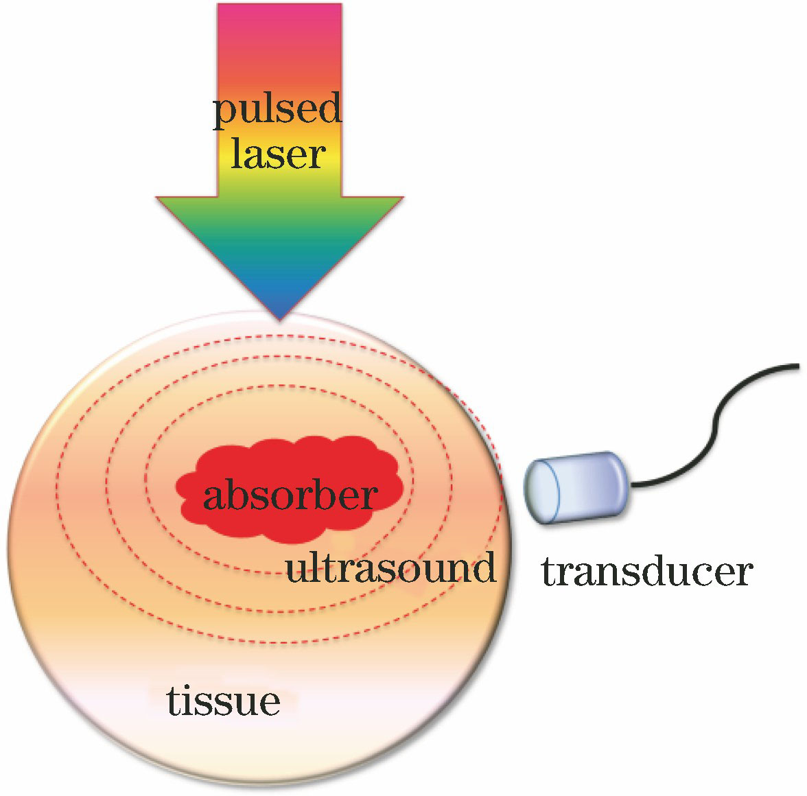
Fig. 1. Principle of photoacoustic imaging
![Photoacoustic imaging systems. (a) Transmission-mode OR-PAM system; (b) reflection-mode OR-PAM system; (c) AR-PAM system with dark-field illumination; (d) PACT system with ring-shaped UTA; (e) PACT system with linear UTA; (f) PACT system with hemispherically shaped UTA; (g) PACT system based on 2D Fabry-Perot interferometer acoustic sensor; (h) side-viewing intravascular PA catheter[46]](/richHtml/lop/2019/56/7/070001/img_2.jpg)
Fig. 2. Photoacoustic imaging systems. (a) Transmission-mode OR-PAM system; (b) reflection-mode OR-PAM system; (c) AR-PAM system with dark-field illumination; (d) PACT system with ring-shaped UTA; (e) PACT system with linear UTA; (f) PACT system with hemispherically shaped UTA; (g) PACT system based on 2D Fabry-Perot interferometer acoustic sensor; (h) side-viewing intravascular PA catheter[46]
Fig. 3. Photoacoustic imaging of breast tumor[30]. (a) UV-PAM image of fixed, unprocessed breast tumor; (b) H&E-stained histologic image of same area shown in Fig. (a) acquired after sectioning and staining breast tissue; (c)(d) zoomed UV-PAM and H&E-stained images of red dashed regions in Fig. (a) and (b), respectively; (e)(f) zoomed UV-PAM and H&E images of yellow dashed regions in Fig. (a) and (b), respectively; (g) zoomed UV-PAM image of orange dashed region in Fig. (a)
Fig. 4. Absorption spectra of hemoglobin[83]
Fig. 5. Oxygenation levels within tumor using photoacoustic imaging. (a) Two single-wavelength PAR amplitude technique; (b) two-wavelength differential PAR amplitude technique; (c) two single-wavelength phase-filtered differential PAR technique; (d) two-wavelength phase-filtered differential PAR technique[80]
Fig. 6. Photoacoustic viscoelastic imaging of tumor. (a) Optical absorption image of tumor; (b) viscoelasticity image of tumor; (c) averaged amplitude and phase delay of PA signal from marked tumor region and normal tissue [91]
Fig. 7. Photoacoustic power spectral imaging of prostate tumors[100]. (a) Ultrasonic imaging in region of interests for tumor and normal tissue; (b) mid-band fitting result after superposition of PA image and ultrasonic image; (c) slope after superposition of PA image and ultrasonic image; (d) intercept after superposition of PA image and ultrasonic image

Set citation alerts for the article
Please enter your email address
© Copyright 2018-2021 | Chinese Laser Press. All Rights Reserved 沪ICP备15018463号-20