• Chinese Journal of Lasers
  • Vol. 50, Issue 15, 1507103 (2023)
Xiaojuan Quan, Chenshuang Zhang, Danying Lin, Bin Yu*, and Junle Qu
Author Affiliations
  • Shenzhen Key Laboratory of Photonics and Biophotonics, Key Laboratory of Optoelectronic Devices and Systems of Ministry of Education and Guangdong Province, College of Physics and Optoelectronic Engineering, Shenzhen University, Shenzhen 518060, Guangdong, China
  • show less
    DOI: 10.3788/CJL230480 Cite this Article Set citation alerts
    Xiaojuan Quan, Chenshuang Zhang, Danying Lin, Bin Yu, Junle Qu. Two-Photon Sub-Diffraction Multifocal Structured Illumination Microscopy[J]. Chinese Journal of Lasers, 2023, 50(15): 1507103 Copy Citation Text show less
    Schematic of an optical system using a DOE
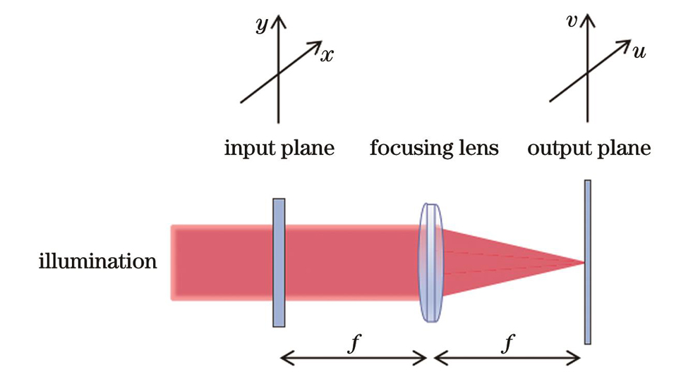
    Fig. 1. Schematic of an optical system using a DOE
    Flow charts of GS algorithm (left) and improved GS algorithm (right)
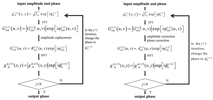
    Fig. 2. Flow charts of GS algorithm (left) and improved GS algorithm (right)
    Schematic of amplitude constraint of the output plane, where the region A of the output plane is divided into 9 large square regions and other regions [amplitude operation (Ⅰ)] and the large square region consists of small square region [amplitude operation (Ⅱ)] and square box 1, square box 2 and square box 3 [amplitude operation (Ⅲ)] from small to large. (a) Amplitude of the output plane; (b) magnified image of the region A in Fig. 3(a)
    Fig. 3. Schematic of amplitude constraint of the output plane, where the region A of the output plane is divided into 9 large square regions and other regions [amplitude operation (Ⅰ)] and the large square region consists of small square region [amplitude operation (Ⅱ)] and square box 1, square box 2 and square box 3 [amplitude operation (Ⅲ)] from small to large. (a) Amplitude of the output plane; (b) magnified image of the region A in Fig. 3(a)
    Results of simulation analysis of normal diffraction limited spot array and sub-diffraction spot array (SSA). (a)(b) Intensity distributions of normal diffraction limited spot arrays and SSA; (c) fitted normalized intensity profiles along the dashed line across a single spot in the central of the normal diffraction limited spot arrays and the SSA; (d) fitted normalized intensity profiles along the dashed line across the three points
    Fig. 4. Results of simulation analysis of normal diffraction limited spot array and sub-diffraction spot array (SSA). (a)(b) Intensity distributions of normal diffraction limited spot arrays and SSA; (c) fitted normalized intensity profiles along the dashed line across a single spot in the central of the normal diffraction limited spot arrays and the SSA; (d) fitted normalized intensity profiles along the dashed line across the three points
    Schematic of 2P-sMSIM system
    Fig. 5. Schematic of 2P-sMSIM system
    Imaging results of normal diffraction limited spot array and SSA. (a)(b) Images of normal diffraction limited spot array and SSA; (c) intensity profiles along the dotted lines in Fig. 6(a) and Fig. 6(b)
    Fig. 6. Imaging results of normal diffraction limited spot array and SSA. (a)(b) Images of normal diffraction limited spot array and SSA; (c) intensity profiles along the dotted lines in Fig. 6(a) and Fig. 6(b)
    2P-sMSIM imaging of microtubules in a BS-C-1 cell. (a) Wide-field image obtained with normal diffraction limited spot array excitation (obtain with overlapping all spot arrays); (b) wide-field image obtained with SSA excitation (obtain with overlapping all spot arrays); (c) 2P-MSIM imaging; (d) 2P-sMSIM imaging; (e) intensity profiles along the dotted lines in Fig. 7(c) and Fig. 7(d)
    Fig. 7. 2P-sMSIM imaging of microtubules in a BS-C-1 cell. (a) Wide-field image obtained with normal diffraction limited spot array excitation (obtain with overlapping all spot arrays); (b) wide-field image obtained with SSA excitation (obtain with overlapping all spot arrays); (c) 2P-MSIM imaging; (d) 2P-sMSIM imaging; (e) intensity profiles along the dotted lines in Fig. 7(c) and Fig. 7(d)
    2P-sMSIM imaging of mitochondria. (a) Wide-field image obtained by using normal diffraction limited spot excitation; (b) wide-field image obtained by using SSA excitation; (c) 2P-MSIM imaging; (d) 2P-sMSIM imaging; (e)(f) magnified view of the rectangle region boxed in Fig. 8(c) and Fig. 8(d)
    Fig. 8. 2P-sMSIM imaging of mitochondria. (a) Wide-field image obtained by using normal diffraction limited spot excitation; (b) wide-field image obtained by using SSA excitation; (c) 2P-MSIM imaging; (d) 2P-sMSIM imaging; (e)(f) magnified view of the rectangle region boxed in Fig. 8(c) and Fig. 8(d)
    Xiaojuan Quan, Chenshuang Zhang, Danying Lin, Bin Yu, Junle Qu. Two-Photon Sub-Diffraction Multifocal Structured Illumination Microscopy[J]. Chinese Journal of Lasers, 2023, 50(15): 1507103
    Download Citation