The multifocal structure light microscope (MSIM) can achieve an imaging depth of 50 μm and a diffraction limited resolution that is two times that of the traditional wide-field structure light technique. However, factors such as fluorescence noise, scattering, and aberration limit its chromatographic capability and imaging depth when conducting super-resolution imaging of thick samples. To overcome these limitations, the MSIM technique combined with two-photon excitation, is implemented to obtain a two-photon multifocus structured light illumination microscope (2P-MSIM), and the imaging depth and resolution of MSIM are thus improved. The MSIM has great application potential in the field of fast super-resolution microscopic imaging of deep tissues in vivo. However, owing to the diffraction limit, the enhancement of the existing 2P-MSIM spatial resolution is limited. To further improve the spatial resolution of 2P-MSIM, a two-photon sub-diffraction multifocal structure illumination microscopy (2P-sMSIM) is proposed in this paper.
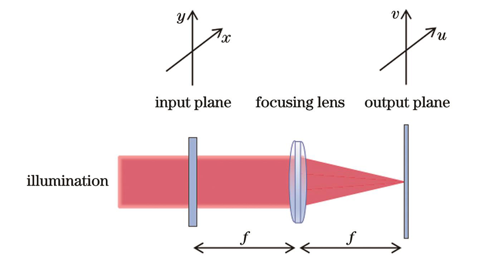
Using the improved Gerchberg-Saxton (GS) phase recovery algorithm, the conditions restricting the amplitude and the phase were enhanced in specific locations on the spectrum plane (the input plane reached the output plane after Fourier transform); subsequently, the amplitude and phase were constrained. After several iterations, the phase diagram of the sub-diffraction spot arrays (SSAs) was calculated, and the initial SSAs were obtained. The sidelobe intensity was controlled at approximately one-tenth of the center intensity (the sidelobe effect can be eliminated by the subsequent image-processing algorithm), and the algorithm parameter optimization was completed to obtain an SSA suitable for system imaging. Raw data for SSA and normal diffraction limited spot arrays were obtained. Next, Gaussian pinhole filtering, pixel relocation, and deconvolution algorithms were used to obtain the SSA and normal diffraction limited spot array images. The resolution of 2P-MSIM imaging was improved under the excitation of the SSA.
The simulation results for the SSA and normal diffraction limited spot arrays are shown in Fig. 4. The average value of the full width at half maximum (FWHM) of the normal diffraction limited spot arrays is approximately 10.60 pixel; the average FWHM for a single focal point in the SSA is approximately 7.64 pixel. As a result, the SSA is 72% of the size of the normal diffraction limited spot arrays, achieving its design purpose. The phase diagram designed above is loaded onto a liquid-crystal spatial light modulator, and the SSA generated by the 2P-sMSIM system is used to excite the uniform eosin solution. Normal diffraction limited spot arrays and SSAs are obtained, and the experimental results are shown in Fig. 6. The normal diffraction limited spot arrays have an average FWHM of 0.71 μm±0.07 μm for a single point, while the SSA has an average FWHM of 0.57 μm±0.07 μm. The SSAs are 80% the size of the normal diffraction limited spot arrays. SSAs can effectively reduce the size of the point diffusion function of the system to further improve the resolution of the 2P-MSIM system. To test the 2P-sMSIM spatial resolution, cell microtubules were used as samples for the experiments, and the results are shown in Fig. 7. the normal diffraction limited spot arrays have a resolution of 151 nm±5 nm after scanning and 135 nm±5 nm after SSA scanning. The image resolution improved by approximately 16 nm after the sub-diffraction-focusing lattice scanning. The experiments prove that the SSA designed using this algorithm can effectively improve the resolution of 2P-MSIM imaging. To further demonstrate the imaging effects of the 2P-sMSIM, commercial mitochondrial sections obtained from Invitrogen were used for fluorescence imaging. The results are presented in Fig. 8. The images after the SSA scan processing are clearer, and the image quality is better. A ring structure that cannot be distinguished in the normal diffraction limited spot array scanned images is clearly observed in Fig. 8(f). The experiments further prove that SSA can effectively improve the resolution and imaging quality of the 2P-MSIM system.
A design method for SSAs is developed in this study and applied to a 2P-MSIM for experimental verification in bioimaging. In the simulation experiment, normal diffraction limited spot arrays are compared with the SSA. By calculating their standard deviations, the SSA can shrink the spots to 72% of the normal diffraction limited spot arrays. The SSA is reduced to 80% of that of the normal diffraction limited spot arrays, proving that the SSA can improve the spatial resolution. Compared with the normal diffraction limited spot arrays, microtubule imaging experiments further verify the effectiveness of the algorithm, and its resolution is improved by approximately 16-135 nm. Mitochondrial imaging experiments also demonstrate the effectiveness and practicability of this algorithm, which lays the foundation for further applications of this algorithm in super-resolution imaging of living cells and tissues.