• Acta Optica Sinica
  • Vol. 39, Issue 2, 0218001 (2019)
Jinyu Li1、*, Youhua Chen1、2、*, Wei Han1, Yu Shang2, and Zhiguo Gui2
Author Affiliations
  • 1 Engineering Technology Research Center of Shanxi Province for Opto-Electronic Information and Instrument, North University of China, Taiyuan, Shanxi 0 30051, China
  • 2 Shanxi Provincial Key Laboratory for Biomedical Imaging and Big Data, North University of China, Taiyuan, Shanxi 0 30051, China
  • show less
    DOI: 10.3788/AOS201939.0218001 Cite this Article Set citation alerts
    Jinyu Li, Youhua Chen, Wei Han, Yu Shang, Zhiguo Gui. Axial Super-Resolution Fluorescence Microscopy Imaging Technology Based on r-ADMM Algorithm[J]. Acta Optica Sinica, 2019, 39(2): 0218001 Copy Citation Text show less
    Experimental setup and principle. (a) Schematic of experimental setup; (b) different penetration depths for different incident angles
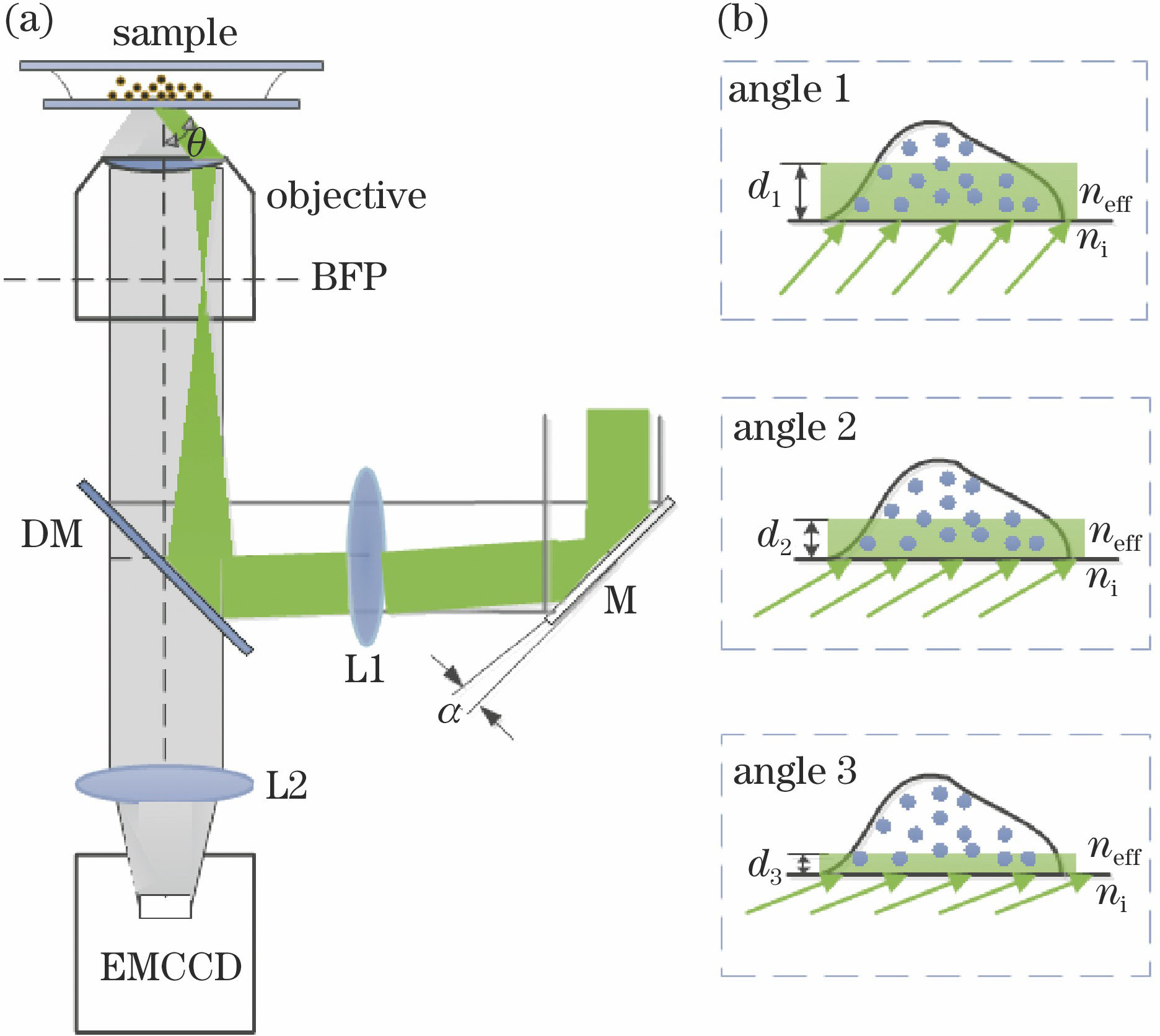
    Fig. 1. Experimental setup and principle. (a) Schematic of experimental setup; (b) different penetration depths for different incident angles
    Flow chart of the algorithm
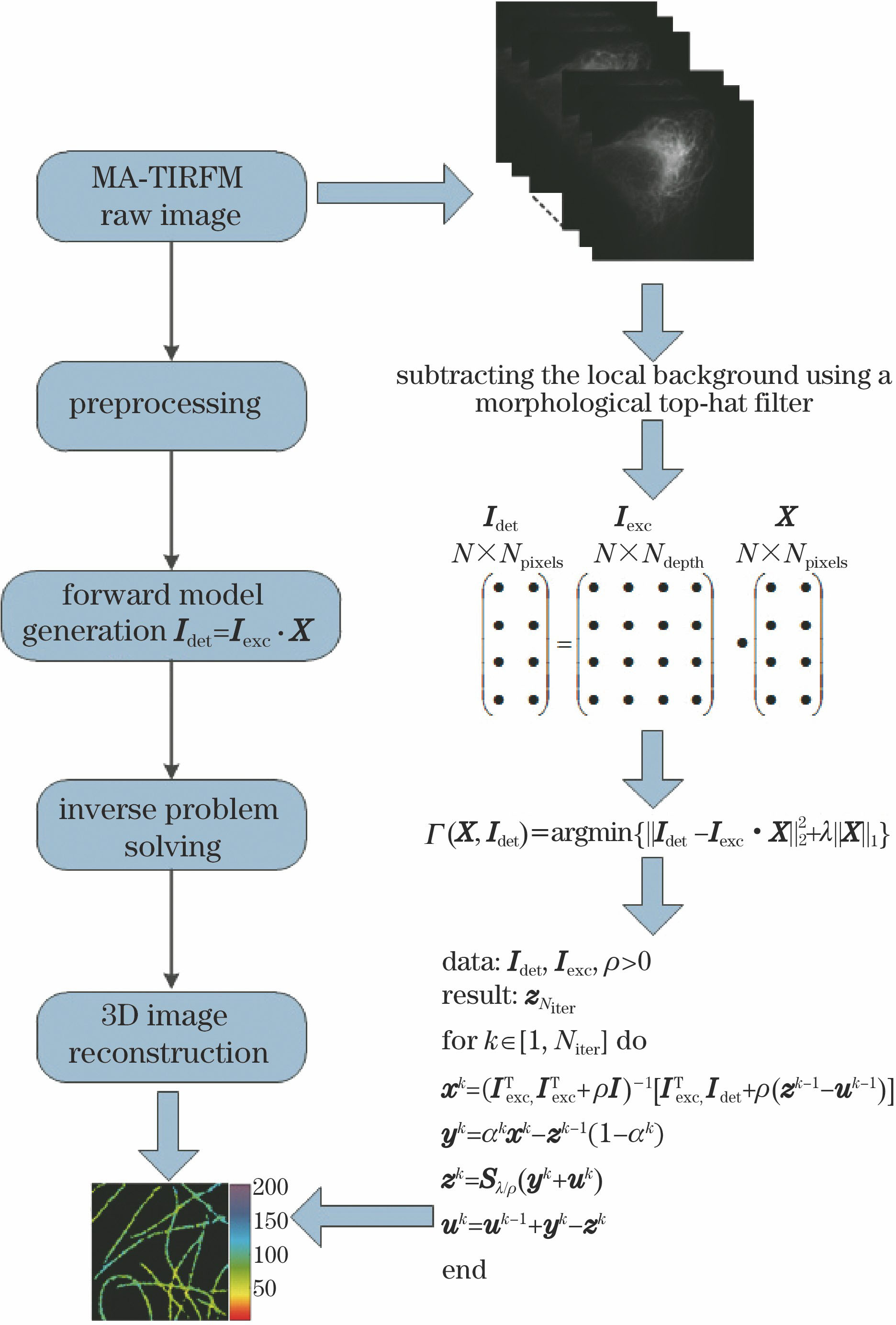
    Fig. 2. Flow chart of the algorithm
    Simulation results of cellular microtubule structures based on MA-TIRFM. (a) Simulated microtubule structures and reconstructed results; (b) accuracy corresponding to different depths at different incident angle numbers
    Fig. 3. Simulation results of cellular microtubule structures based on MA-TIRFM. (a) Simulated microtubule structures and reconstructed results; (b) accuracy corresponding to different depths at different incident angle numbers
    MA-TIRFM imaging of fixed microtubules. (a) Wide-field image of microtubules; (b) three-dimensional map reconstructed by traditional ADMM algorithm; (c) three-dimension map reconstructed by r-ADMM algorithm; (d) 40 nm axial resolution achieved by traditional ADMM algorithm [corresponding to red dot pointed by white arrow in (b)]; (e) 40 nm axial resolution achieved by r-ADMM algorithm [corresponding to red dot pointed by white arrow in (c)]; (f) images of microtubules at different depths reconst
    Fig. 4. MA-TIRFM imaging of fixed microtubules. (a) Wide-field image of microtubules; (b) three-dimensional map reconstructed by traditional ADMM algorithm; (c) three-dimension map reconstructed by r-ADMM algorithm; (d) 40 nm axial resolution achieved by traditional ADMM algorithm [corresponding to red dot pointed by white arrow in (b)]; (e) 40 nm axial resolution achieved by r-ADMM algorithm [corresponding to red dot pointed by white arrow in (c)]; (f) images of microtubules at different depths reconst
    Comparison chart of residual error curves. (a) Residual error versus iterations for ADMM and r-ADMM algorithms; (b) residual error versus iterations for r-ADMM algorithm with different r values
    Fig. 5. Comparison chart of residual error curves. (a) Residual error versus iterations for ADMM and r-ADMM algorithms; (b) residual error versus iterations for r-ADMM algorithm with different r values
    Dynamic process of active mitochondria. (a) Wide-field image and reconstructed map; (b) fission (red triangle) andfusion (blue triangle) events in dotted box in Fig. 6 (a) captured by MA-TIRFM image stacks for 2 s time interval
    Fig. 6. Dynamic process of active mitochondria. (a) Wide-field image and reconstructed map; (b) fission (red triangle) andfusion (blue triangle) events in dotted box in Fig. 6 (a) captured by MA-TIRFM image stacks for 2 s time interval
    AlgorithmIterations under different ρ valuesTime /s
    ρ=0.3ρ=0.4ρ=0.5
    Traditional ADMM algorithm24191716
    r-ADMM algorithm16151312
    Increase rate of iterative convergence speed /%33.3321.0523.5225
    Table 1. Iterations and time for r-ADMM and traditional ADMM algorithms at different ρ values
    Jinyu Li, Youhua Chen, Wei Han, Yu Shang, Zhiguo Gui. Axial Super-Resolution Fluorescence Microscopy Imaging Technology Based on r-ADMM Algorithm[J]. Acta Optica Sinica, 2019, 39(2): 0218001
    Download Citation