• Spectroscopy and Spectral Analysis
  • Vol. 41, Issue 11, 3468 (2021)
Zhen RUAN1、1;, Peng-fei ZHU3、3;, Lei ZHANG3、3;, Rong-ze CHEN3、3;, Xun-rong LI3、3;, Xiao-ting FU3、3;, Zheng-gu HUANG4、4;, Gang ZHOU4、4;, Yue-tong JI5、5;, and Pu LIAO1、1; 2; *;
Author Affiliations
  • 11. Key Laboratory of Diagnostic Medicine, Ministry of Education; School of Laboratory Medicine, Chongqing Medical University, Chongqing 400016, China
  • 33. Single-Cell Center, Qingdao Institute of Bioenergy and Bioprocess Technology, Chinese Academy of Sciences, Qingdao 266101, China
  • 44. Center Lab, Chongqing Public Health Medical Center, Chongqing 400036, China
  • 55. Qingdao Single-Cell Biotechnology Co., Ltd., Qingdao 266101, China
  • show less
    DOI: 10.3964/j.issn.1000-0593(2021)11-3468-06 Cite this Article
    Zhen RUAN, Peng-fei ZHU, Lei ZHANG, Rong-ze CHEN, Xun-rong LI, Xiao-ting FU, Zheng-gu HUANG, Gang ZHOU, Yue-tong JI, Pu LIAO. Study on Identification of Non-Tuberculosis Mycobacteria Based on Single-Cell Raman Spectroscopy[J]. Spectroscopy and Spectral Analysis, 2021, 41(11): 3468 Copy Citation Text show less
    Original average Raman spectra and difference peaks of six NTMs
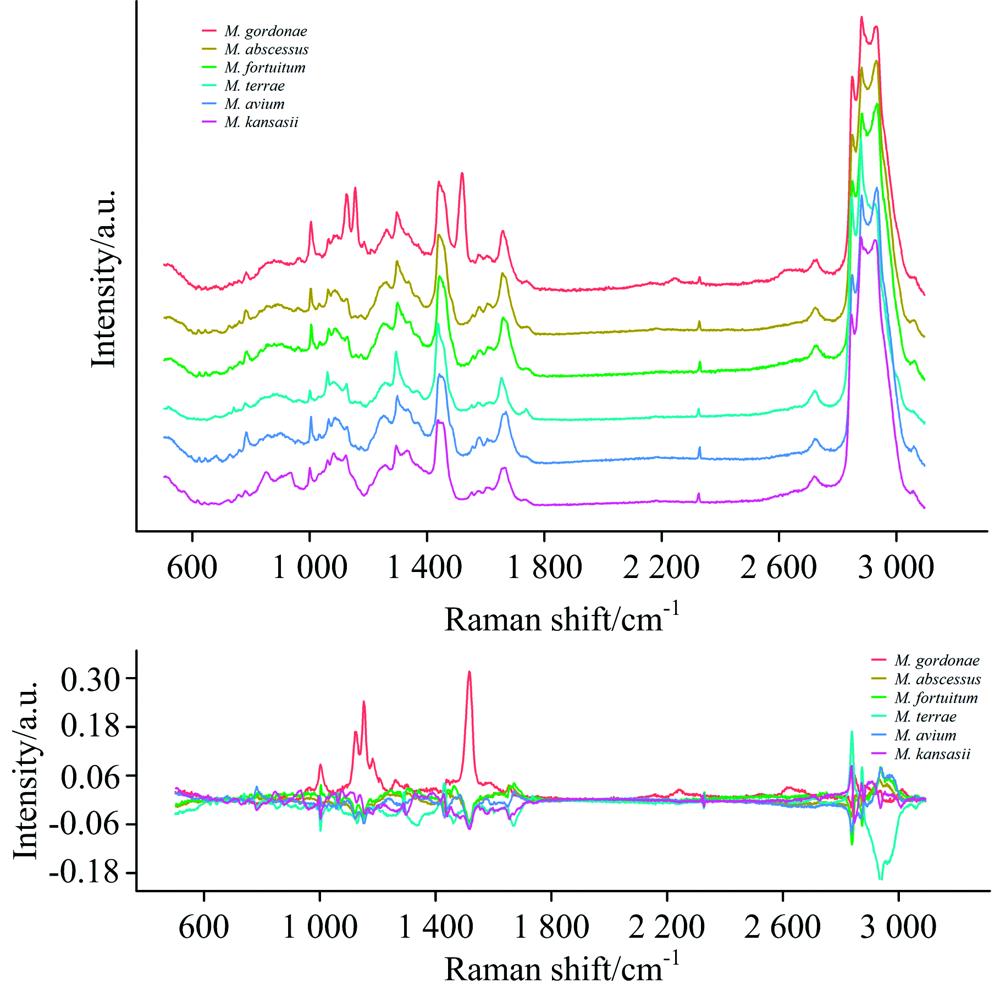
    Fig. 1. Original average Raman spectra and difference peaks of six NTMs
    Pretreatment average Raman spectra of six NTMs
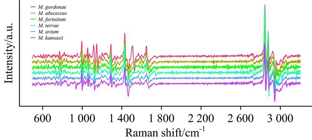
    Fig. 2. Pretreatment average Raman spectra of six NTMs
    t-SNE results of six NTMs SCRS
    Fig. 3. t-SNE results of six NTMs SCRS
    细菌名称光谱数
    脓肿分枝杆菌Mycobacterium abscessus92
    戈登分枝杆菌Mycobacterium gordonae99
    偶发分枝杆菌Mycobacterium fortuitum67
    土分枝杆菌Mycobacterium terrae73
    鸟分枝杆菌Mycobacterium avium68
    堪萨斯分枝杆菌Mycobacterium kansasii154
    Table 1. List of species and the number of Raman spectra from single cells
    Peak position
    /cm-1
    ProteinsLipidsNucleic acidsCarbohydrates
    644.126C—S stretching & C—C twisting of proteins
    726.371Characteristic for phospholipids
    783.39U, T, C
    829.318tyrosine (proteins), O—P—O stretch DNA
    854.267C—O—C skeletal mode of α-anomers
    1 004.49Phenylalanine ring breath
    1 030.24ν(CO), ν(CC), ν(CCO)
    1 084.69Typical phospholipids
    1 128.03C—O stretching
    1 153.82C—C/C—N stretching
    1 172.2C—H in-plane bending mode of tyrosine, (CH) phenylalanine
    1 211.05ν(C-C6H5), tryptophan, phenylalanine
    1 248.78Amide Ⅲ
    1 340.43CH stretching of adenine
    1 449.47CH2 bending and scissoring modes of phospholipids
    1 518.22v(C=C) diagnostic for the presence of a carotenoid structure, most likely a cellular pigment v(C=C) carotenoid
    1 574.87Phenylalanine
    1 604.68Phenylalanine, tyrosine, C=C
    1 669.75Allyl C=C stretches
    Table 2. Molecular structure and composition of Raman peaks
    ModelTest dataset
    SampleCorrectAccuracy/%
    SVM16416399.4
    K-NN16411369.1
    PLS-DA16413985.0
    RF16415695.2
    LDA16416298.8
    XGB16414387.3
    Table 3. Results of six identification models
    SVMPrediction
    M. abscessusM. gordonaeM. fortuitumM. terraeM. aviumM. kansasii
    M. abscessus2300000
    M. gordonae0270000
    M. fortuitum0016000
    M. terrae0002900
    M. avium0000210
    M. kansasii0001048
    Table 4. Prediction results of SVM confusion matrix
    LDAPrediction
    M. abscessusM. gordonaeM. fortuitumM. terraeM. aviumM. kansasii
    M. abscessus2201000
    M. gordonae0260100
    M. fortuitum0016000
    M. terrae0002900
    M. avium0000210
    M. kansasii0000049
    Table 5. Prediction results of LDA confusion matrix
    Zhen RUAN, Peng-fei ZHU, Lei ZHANG, Rong-ze CHEN, Xun-rong LI, Xiao-ting FU, Zheng-gu HUANG, Gang ZHOU, Yue-tong JI, Pu LIAO. Study on Identification of Non-Tuberculosis Mycobacteria Based on Single-Cell Raman Spectroscopy[J]. Spectroscopy and Spectral Analysis, 2021, 41(11): 3468
    Download Citation