• Chinese Optics Letters
  • Vol. 18, Issue 12, 121703 (2020)
Fangyu Wang1、2、3、4, Yuhao Yuan4, Qiang Sun1, Ming Dai1, Li Ai5、6, and Fake Lu4、*
Author Affiliations
  • 1Changchun Institute of Optics, Fine Mechanics and Physics, Chinese Academy of Sciences, Changchun 130033, China
  • 2University of Chinese Academy of Sciences, Beijing 100049, China
  • 3Jilin Yataizhongke Medical Equipment Engineering Technology Research Institute Holding Co., Ltd., Changchun 130000, China
  • 4Department of Biomedical Engineering, Binghamton University, State University of New York, Binghamton, NY 13902, USA
  • 5Jilin Provincial Key Laboratory of Photoelectric Equipment and Instrument Advanced Manufacture Technology, Changchun 130033, China
  • 6Changchun UP Optotech Holding Co., Ltd., Changchun 130033, China
  • show less
    DOI: 10.3788/COL202018.121703 Cite this Article Set citation alerts
    Fangyu Wang, Yuhao Yuan, Qiang Sun, Ming Dai, Li Ai, Fake Lu. Design and implementation of the galvanometer scanning system for reflectance confocal and stimulated Raman scattering microscopy[J]. Chinese Optics Letters, 2020, 18(12): 121703 Copy Citation Text show less
    Optical path layout of the RCM system. APD, avalanche photodiode; PBS, polarizing beam splitter; QWP, quarter-wave plate; RCM, reflectance confocal microscope.
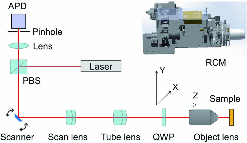
    Fig. 1. Optical path layout of the RCM system. APD, avalanche photodiode; PBS, polarizing beam splitter; QWP, quarter-wave plate; RCM, reflectance confocal microscope.
    Electrical control and data acquisition diagram of the RCM.
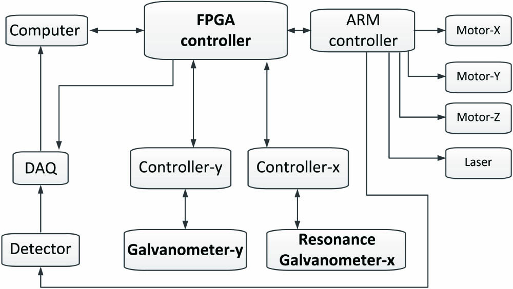
    Fig. 2. Electrical control and data acquisition diagram of the RCM.
    Control principle diagram in the RCM system.
    Fig. 3. Control principle diagram in the RCM system.
    FPGA controller realized synchronization of galvanometer scanning, data acquisition, and image formation.
    Fig. 4. FPGA controller realized synchronization of galvanometer scanning, data acquisition, and image formation.
    Demonstration of in vivo video-rate imaging of human skin using the RCM system we designed. Scale bar, 150 μm; frame rate, 11 fps.
    Fig. 5. Demonstration of in vivo video-rate imaging of human skin using the RCM system we designed. Scale bar, 150 μm; frame rate, 11 fps.
    Optical path of the SRS microscope. EOM, electro-optic modulator; HWP, half-wave plate.
    Fig. 6. Optical path of the SRS microscope. EOM, electro-optic modulator; HWP, half-wave plate.
    Electrical and data acquisition diagram of the SRS microscope.
    Fig. 7. Electrical and data acquisition diagram of the SRS microscope.
    SRS images of live SKOV-3 ovarian cancer cells at 2854 cm−1 (left, CH2; lipids) and 2940 cm−1 (CH3, proteins). The cell line was purchased from American Type Culture Collection (ATCC) and cultured in McCoy’s 5A Medium (ATCC) with 10% fetal bovine serum (ATCC) for 24 h on a glass coverslip before imaging. The images were acquired with 1024×1024 pixels, and the dwell time of each pixel was 10 μs.
    Fig. 8. SRS images of live SKOV-3 ovarian cancer cells at 2854cm1 (left, CH2; lipids) and 2940cm1 (CH3, proteins). The cell line was purchased from American Type Culture Collection (ATCC) and cultured in McCoy’s 5A Medium (ATCC) with 10% fetal bovine serum (ATCC) for 24 h on a glass coverslip before imaging. The images were acquired with 1024×1024pixels, and the dwell time of each pixel was 10 μs.
    Fangyu Wang, Yuhao Yuan, Qiang Sun, Ming Dai, Li Ai, Fake Lu. Design and implementation of the galvanometer scanning system for reflectance confocal and stimulated Raman scattering microscopy[J]. Chinese Optics Letters, 2020, 18(12): 121703
    Download Citation