• Laser & Optoelectronics Progress
  • Vol. 57, Issue 23, 231701 (2020)
Qi Lin, Yu Lin, Yunde Xie, and Zhongxiong Ma*
Author Affiliations
  • Department of Oral Mucosa, the Affiliated Stomatological Hospital, Fujian Medical University, Fujian Key Laboratory of Oral Diseases, Fuzhou, Fujian 350002, China
  • show less
    DOI: 10.3788/LOP57.231701 Cite this Article Set citation alerts
    Qi Lin, Yu Lin, Yunde Xie, Zhongxiong Ma. Study on Crystal Structure Change of Laser-Ablated Human Dentin Surface Based on X-Ray Diffraction[J]. Laser & Optoelectronics Progress, 2020, 57(23): 231701 Copy Citation Text show less
    XRD pattern of dentin (dash line) and simulated XRD pattern of hydroxyapatite from ICSD-56312 (solid line)
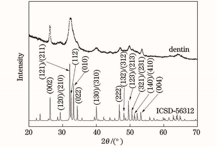
    Fig. 1. XRD pattern of dentin (dash line) and simulated XRD pattern of hydroxyapatite from ICSD-56312 (solid line)
    XRD patterns of dentin before (dash line) and after (solid line) laser irradiation with different energy densities
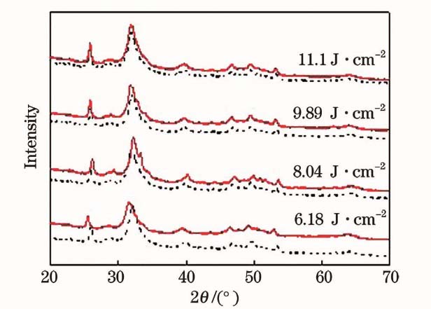
    Fig. 2. XRD patterns of dentin before (dash line) and after (solid line) laser irradiation with different energy densities
    Crystallographically refined HA crystallite shapes after Er,Cr∶YSGG laser irradiation using Popa model
    Fig. 3. Crystallographically refined HA crystallite shapes after Er,Cr∶YSGG laser irradiation using Popa model
    SEM images of dentin before and after laser irradiation (N: dentin before laser irradiation, L: dentin after laser irradiation). (a) Before laser irradiation; (b) after laser irradiation with energy density of 6.18 J/cm2; (c) after laser irradiation with energy density of 8.04 J/cm2; (d) after laser irradiation with energy density of 9.89 J/cm2; (e) after laser irradiation with energy density of 11.1 J/cm2
    Fig. 4. SEM images of dentin before and after laser irradiation (N: dentin before laser irradiation, L: dentin after laser irradiation). (a) Before laser irradiation; (b) after laser irradiation with energy density of 6.18 J/cm2; (c) after laser irradiation with energy density of 8.04 J/cm2; (d) after laser irradiation with energy density of 9.89 J/cm2; (e) after laser irradiation with energy density of 11.1 J/cm2
    Pulse frequency /HzSpot diameter /μmLaser power(machine parameter) /WLaser energy density(experimental parameter) /(J·cm-2)
    2075026.18
    2.58.04
    39.89
    3.511.1
    Table 1. Er,Cr∶YSGG laser parameters
    Laser energy density /(J·cm-2)(002) axial size/nm(130) axial size /nm
    BeforeAfterBeforeAfter
    6.1832.80±4.930.25±4.49.41±3.98.31±4.2
    8.0425.19±5.222.90±5.810.85±2.48.2±4.1
    9.8932.08±4.729.68±4.19.53±2.77.58±3.5
    11.127.70±4.522.60±6.010.12±2.69.13±2.9
    Table 2. Average particle sizes of HA nanocrystals before and after Er, Cr∶YSGG laser irradiation at (002) and (130) positions
    Laser energy density/(J·cm-2)Lattice parameter a/nmLattice parameter c/nm
    BeforeAfterBeforeAfter
    6.180.9450.9440.6880.689
    8.040.9440.9430.6880.688
    9.890.9440.9450.6880.689
    11.10.9450.9440.6880.689
    Table 3. Lattice parameters of nano HA in dentin before and after Er,Cr∶YSGG laser irradiation
    Qi Lin, Yu Lin, Yunde Xie, Zhongxiong Ma. Study on Crystal Structure Change of Laser-Ablated Human Dentin Surface Based on X-Ray Diffraction[J]. Laser & Optoelectronics Progress, 2020, 57(23): 231701
    Download Citation