• Chinese Journal of Lasers
  • Vol. 50, Issue 3, 0307108 (2023)
Lin Yao1, Chenyang Yu1, Kaiyuan Liu1, Xiaofeng Deng1, Zhihua Ding1, and Peng Li1、2、*
Author Affiliations
  • 1College of Optical Science and Engineering, Zhejiang University, Hangzhou 310027, Zhejiang, China
  • 2Intelligent Optics & Photonics Research Center Jiaxing Research Institute, Zhejiang University, Jiaxing 324000, Zhejiang, China
  • show less
    DOI: 10.3788/CJL221230 Cite this Article Set citation alerts
    Lin Yao, Chenyang Yu, Kaiyuan Liu, Xiaofeng Deng, Zhihua Ding, Peng Li. Three‐Dimensional Defects Inspection of Bioprosthetic Valves[J]. Chinese Journal of Lasers, 2023, 50(3): 0307108 Copy Citation Text show less
    Schematic of SS-OCT system
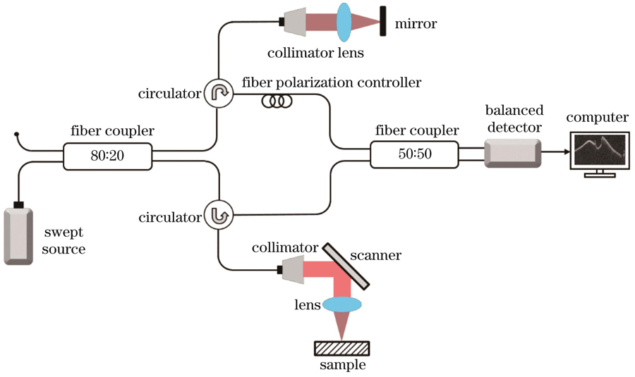
    Fig. 1. Schematic of SS-OCT system
    OCT structural imaging results of bioprosthetic valves-tricuspid valve (scale bar: 2 mm). (a) Schematic diagram of tricuspid valve stent; (b) three-dimensional OCT structure (Z-X-Y); (c) en-face (X-Y) image of three-dimensional OCT structure; (d) OCT structural cross-section, corresponding to the position of the dashed line in figure (c)
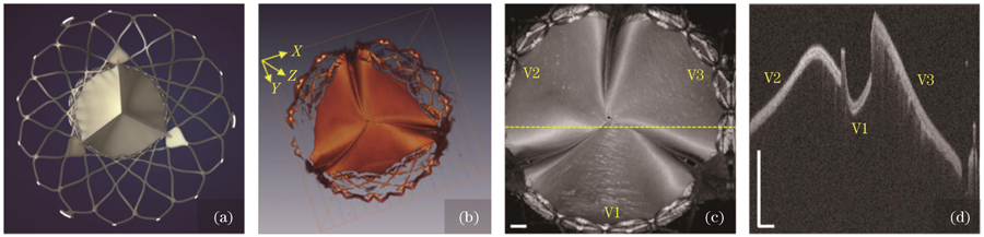
    Fig. 2. OCT structural imaging results of bioprosthetic valves-tricuspid valve (scale bar: 2 mm). (a) Schematic diagram of tricuspid valve stent; (b) three-dimensional OCT structure (Z-X-Y); (c) en-face (X-Y) image of three-dimensional OCT structure; (d) OCT structural cross-section, corresponding to the position of the dashed line in figure (c)
    OCT structural imaging results of fiber surface of valve leaflets made of swine heart pericardium (scale bar: 2 mm). (a)(c) OCT en-face images of fiber surface in normal valve leaflets; (b)(d) two-dimensional OCT structural cross-sections, corresponding to the positions of the dashed line in figures (a) and (c); (e)(g) OCT en-face images of fiber surface in abnormal valve leaflets; (f)(h) two-dimensional OCT structural cross-sections, corresponding to the positions of the dashed line in figures (e) and (g), where the arrows point the obvious abnormal regions
    Fig. 3. OCT structural imaging results of fiber surface of valve leaflets made of swine heart pericardium (scale bar: 2 mm). (a)(c) OCT en-face images of fiber surface in normal valve leaflets; (b)(d) two-dimensional OCT structural cross-sections, corresponding to the positions of the dashed line in figures (a) and (c); (e)(g) OCT en-face images of fiber surface in abnormal valve leaflets; (f)(h) two-dimensional OCT structural cross-sections, corresponding to the positions of the dashed line in figures (e) and (g), where the arrows point the obvious abnormal regions
    OCT structural imaging results of smooth layer and interlayer defects and cutting defects of valve leaflets made of swine heart pericardium (scale bar: 2 mm). (a)(c) OCT en-face images of damaged smooth layer and wrinkled smooth layer in abnormal valve leaflets; (b)(d) two-dimensional OCT structural cross-sections, corresponding to the positions of the dashed line in figures (a) and (c); (e)(g) OCT en-face images of abnormal valve leaflets with interlayer defects and cutting defects; (f)(h) two-dimensional OCT structural cross-sections, corresponding to the positions of the dashed line in figures (e) and (g), the arrows point the obvious abnormal regions
    Fig. 4. OCT structural imaging results of smooth layer and interlayer defects and cutting defects of valve leaflets made of swine heart pericardium (scale bar: 2 mm). (a)(c) OCT en-face images of damaged smooth layer and wrinkled smooth layer in abnormal valve leaflets; (b)(d) two-dimensional OCT structural cross-sections, corresponding to the positions of the dashed line in figures (a) and (c); (e)(g) OCT en-face images of abnormal valve leaflets with interlayer defects and cutting defects; (f)(h) two-dimensional OCT structural cross-sections, corresponding to the positions of the dashed line in figures (e) and (g), the arrows point the obvious abnormal regions
    Lin Yao, Chenyang Yu, Kaiyuan Liu, Xiaofeng Deng, Zhihua Ding, Peng Li. Three‐Dimensional Defects Inspection of Bioprosthetic Valves[J]. Chinese Journal of Lasers, 2023, 50(3): 0307108
    Download Citation