Heart valve disease is a growing public health concern worldwide. A prosthetic heart valve is a heart implant intervention medical device for the treatment of heart valve disease, which mainly includes bioprosthetic and mechanical valves. Diseased native valves are often replaced with bioprosthetic valves made from porcine or bovine pericardium, which has a lower risk of thrombosis and hemodynamic advantages than mechanical valves. Nonetheless, bioprosthetic valves do not have long-term durability, mainly because of their early structural failure. Therefore, an in vitro fatigue test is required for manufactured bioprosthetic valves; further, it is very important to evaluate the quality of the valve after the fatigue test, thereby obtaining a basis for the optimization of valve performance.
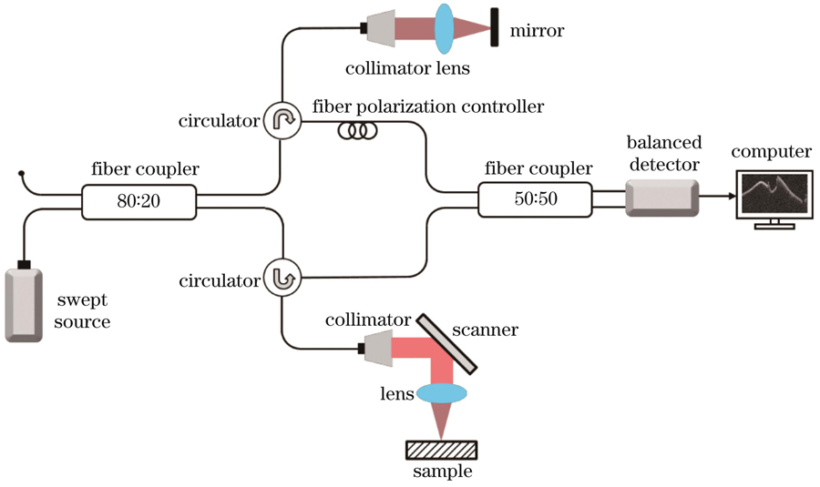
The OCT light source was a MEMS-tunable vertical cavity surface-emitting laser (VCSEL, Thorlabs, SL131090). The laser could sweep at a rate of 100 kHz over a broad spectral bandwidth of ~100 nm with a center wavelength of 1300 nm, providing an experimental axial resolution of ~16 μm and an imaging depth of ~11 mm in air. The output light from the laser source was first fiber-coupled into an interferometer, where the light was split by an 80:20 fiber coupler into a sample arm and reference arm. In the OCT sample arm, a scanning lens (Thorlabs, LSM05) with an effective focal length of 54 mm was used to collimate the detection light on the sample, providing an experimental lateral resolution of ~32 μm, and an X-Y galvanometer was adopted for three-dimensional (3D) volume scanning. The light backscattered from the sample was recombined with the light reflected from the reference mirror, and the interference signal was detected using a balanced detector (Thorlabs, PDB470C). A stepwise raster scanning protocol (Z-X-Y) was used for volumetric imaging, with 1000 A-lines per B-frame (fast-scan, X-direction) and 1000 B-frames at 1000 tomographic positions per volume (slow-scan, Y-direction). OCT imaging covered a field of view (FOV) of 12 mm (X)×12 mm (Y) of the swine heart valve leaflets, and a wide FOV of 28.5 mm (X)×28.5 mm (Y) of the bioprosthetic valves. The captured interference data were converted to amplitude form using a fast Fourier transformation (FFT) processed on the MATLAB (MathWorks) platform. The bioprosthetic valve surface boundary fitting algorithm transforms the depth coordinates of the bioprosthetic valve amplitude structure according to the fitting results, such that the overall trend of the surface boundary is smoothened, but the high-frequency changes in fiber bundles and abnormal protrusions are preserved. The OCT amplitude images were then displayed as a 3D (Z-X-Y) structure view and an en-face (X-Y) maximum intensity projection (MIP) of the 3D structure.
The main advantage of OCT is its ability to acquire large field-of-view two-dimensional (2D) tomograms and 3D volume data. From the structural diagram, it can be concluded that the abnormal direction of the fiber bundles on the surface of the valve leaflet fiber layer (see Fig. 3), damaged and folded surface of the smooth layer (see Fig. 4), abnormal defects between the layers (Fig. 4), and cutting defects are valuable information which are suitable for the inspection of valve leaflet defects.
This paper proposes a 3D defect inspection method for bioprosthetic valves based on OCT technology, which can achieve high-resolution, large field of view, and real-time 3D structural imaging. The method is used to perform 3D imaging on the complete bioprosthetic valve stent and valve leaflets and realize the abnormal detection of the fiber layer, smooth layer, interlayer defects, and cutting defects. The obtained results show that the method can realize high-resolution three-dimensional defect inspection of bioprosthetic valves, which is helpful for biological scientists in evaluating valve quality. Further, the method can be used in the field of valve manufacturing and inspection.