• Acta Optica Sinica
  • Vol. 41, Issue 17, 1726001 (2021)
Yizhe Yang1, Zhongfa Liu1, Yu Fang1, Xiaojing Wu2、*, Yong Yang1、**, and Siwei Zhu2
Author Affiliations
  • 1Institute of Modern Optics, Nankai University, Tianjin 300071, China
  • 2Tianjin Union Medical Centre, Tianjin 300121, China
  • show less
    DOI: 10.3788/AOS202141.1726001 Cite this Article Set citation alerts
    Yizhe Yang, Zhongfa Liu, Yu Fang, Xiaojing Wu, Yong Yang, Siwei Zhu. Influencing Factors and Improved Methods of Spatial Resolution of Focused Refractive Index Microscopy Imaging System[J]. Acta Optica Sinica, 2021, 41(17): 1726001 Copy Citation Text show less
    Polarization selective absorption properties of graphene under total internal reflection conditions. (a) Schematic of sandwich structure containing graphene; (b) reflectivity curves of TE polarized light and TM polarized light
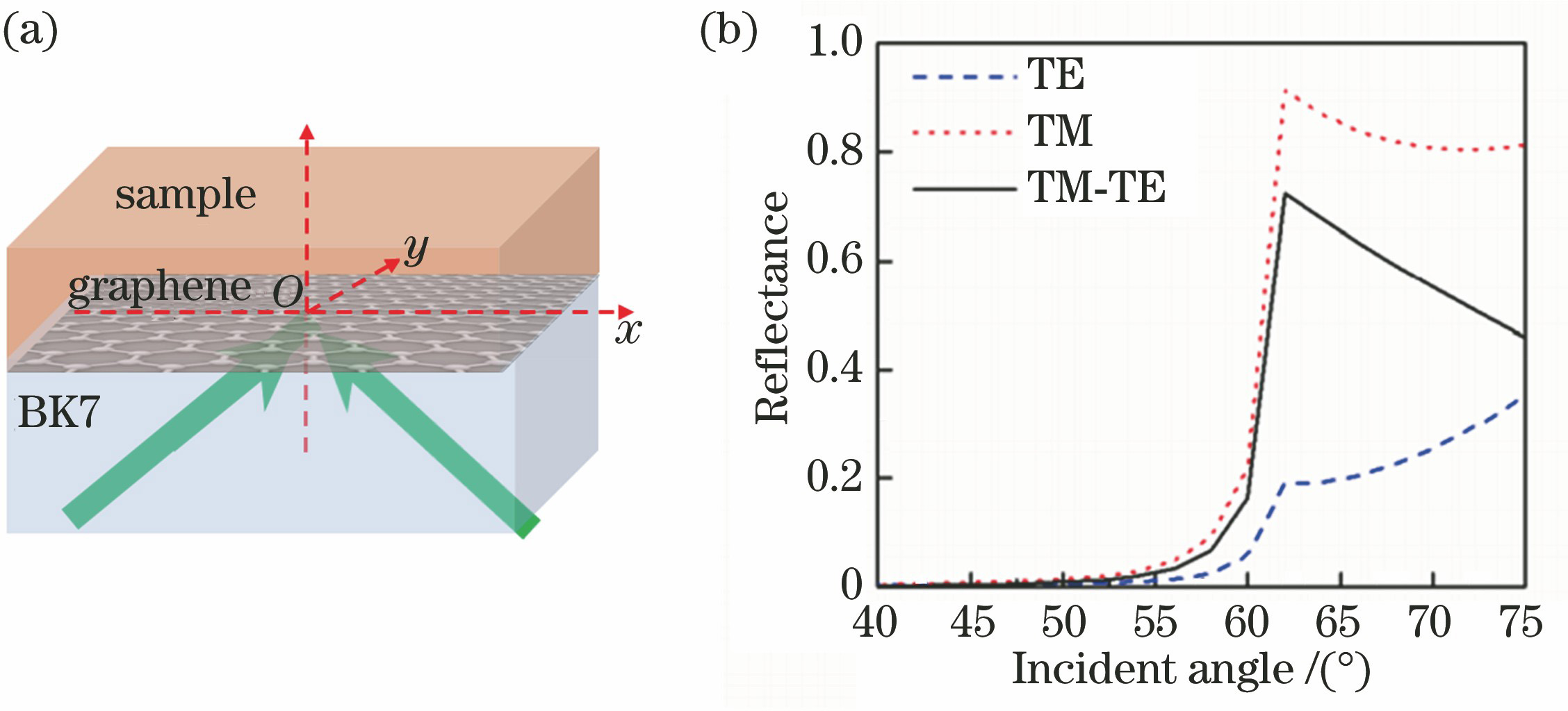
    Fig. 1. Polarization selective absorption properties of graphene under total internal reflection conditions. (a) Schematic of sandwich structure containing graphene; (b) reflectivity curves of TE polarized light and TM polarized light
    45° generalized column vector incident light focusing. (a) Polarization schematic diagram; (b) intensity of longitudinal electric field component; (c) intensity of radial electric field component; (d) intensity of angular electric field component; (e) width at diameter of radial electric field beam; (f) width at diameter of angular electric field beam
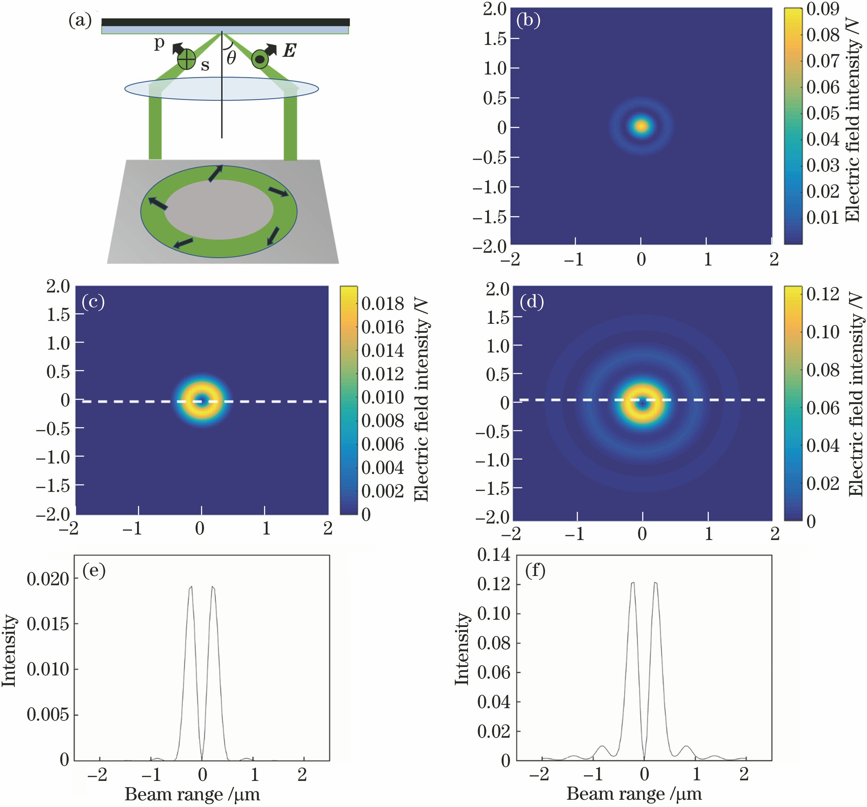
    Fig. 2. 45° generalized column vector incident light focusing. (a) Polarization schematic diagram; (b) intensity of longitudinal electric field component; (c) intensity of radial electric field component; (d) intensity of angular electric field component; (e) width at diameter of radial electric field beam; (f) width at diameter of angular electric field beam
    Model of slim beam scanning multilayer medium
    Fig. 3. Model of slim beam scanning multilayer medium
    Model of ideal slim beam scanning PS medium ball
    Fig. 4. Model of ideal slim beam scanning PS medium ball
    Model of 45° generalized cylindrical vector beam scanning PS medium ball
    Fig. 5. Model of 45° generalized cylindrical vector beam scanning PS medium ball
    Refractive index curves measured at diameter of ball scanned by different beams. (a) Refractive index curve measured at diameter of ball scanned by slim beam; (b) refractive index curve measured at diameter of ball scanned by 45° generalized cylindrical vector beam
    Fig. 6. Refractive index curves measured at diameter of ball scanned by different beams. (a) Refractive index curve measured at diameter of ball scanned by slim beam; (b) refractive index curve measured at diameter of ball scanned by 45° generalized cylindrical vector beam
    Deconvolution and theoretical simulation results. (a) Simulation results of ideal slim beam and generalized column vector beam sweeping over small ball 2; (b) result of ball 2 after eliminating influence of wide beam
    Fig. 7. Deconvolution and theoretical simulation results. (a) Simulation results of ideal slim beam and generalized column vector beam sweeping over small ball 2; (b) result of ball 2 after eliminating influence of wide beam
    Optical path of focused graphene sensor imaging
    Fig. 8. Optical path of focused graphene sensor imaging
    Characterization diagram of PS beads. (a) Microscope characterization diagram; (b) Coulter characterization diagram
    Fig. 9. Characterization diagram of PS beads. (a) Microscope characterization diagram; (b) Coulter characterization diagram
    Result of optical system scanning 1st PS ball. (a) Bright field diagram under optical system; (b) refractive index distribution obtained by scanning 1st PS ball
    Fig. 10. Result of optical system scanning 1st PS ball. (a) Bright field diagram under optical system; (b) refractive index distribution obtained by scanning 1st PS ball
    Deconvolution results of ball experiment. (a) Refractive index distribution obtained by scanning 2nd PS ball by optical system; (b) refractive index distribution of PS ball after one-dimensional transverse deconvolution; (c) refractive index distribution of PS ball after two-dimensional transverse and longitudinal deconvolution; (d) refractive index curves measured at diameter of PS ball recovered by deconvolution
    Fig. 11. Deconvolution results of ball experiment. (a) Refractive index distribution obtained by scanning 2nd PS ball by optical system; (b) refractive index distribution of PS ball after one-dimensional transverse deconvolution; (c) refractive index distribution of PS ball after two-dimensional transverse and longitudinal deconvolution; (d) refractive index curves measured at diameter of PS ball recovered by deconvolution
    Yizhe Yang, Zhongfa Liu, Yu Fang, Xiaojing Wu, Yong Yang, Siwei Zhu. Influencing Factors and Improved Methods of Spatial Resolution of Focused Refractive Index Microscopy Imaging System[J]. Acta Optica Sinica, 2021, 41(17): 1726001
    Download Citation