• Laser & Optoelectronics Progress
  • Vol. 58, Issue 14, 1417001 (2021)
Yiming Liu and Zhiyong Xiao*
Author Affiliations
  • School of Artificial Intelligence and Computer Science, Jiangnan University, Wuxi, Jiangsu 214122, China
  • show less
    DOI: 10.3788/LOP202158.1417001 Cite this Article Set citation alerts
    Yiming Liu, Zhiyong Xiao. Automatic Segmentation Algorithm of Liver Tumor Based on Feature Fusion[J]. Laser & Optoelectronics Progress, 2021, 58(14): 1417001 Copy Citation Text show less
    Structure of deeply-supervised net
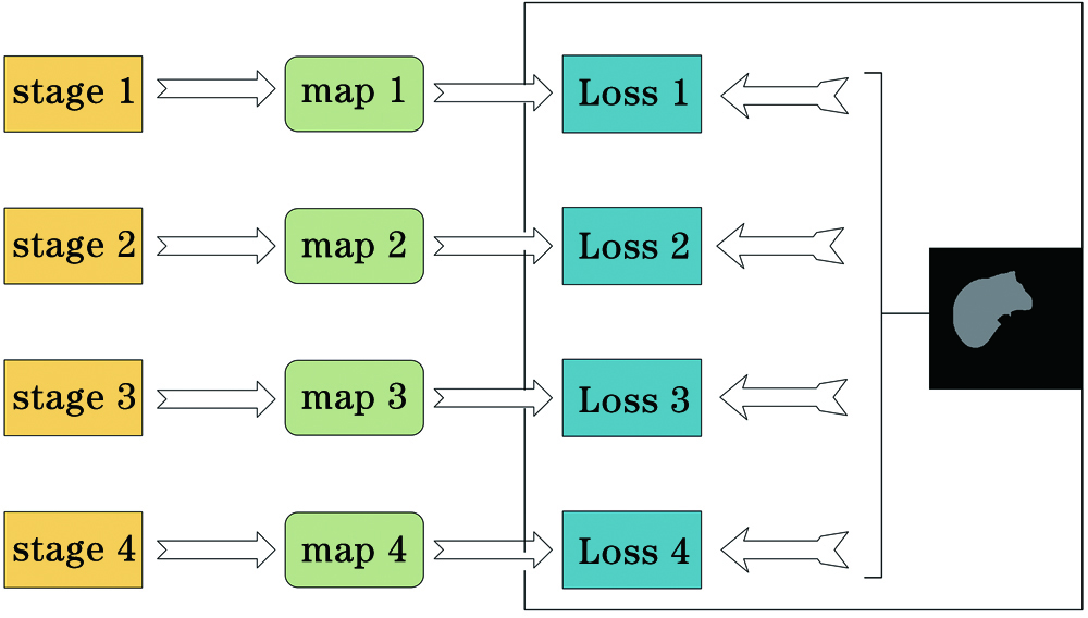
    Fig. 1. Structure of deeply-supervised net
    Flow chart of our experiment
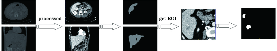
    Fig. 2. Flow chart of our experiment
    Network structure proposed in this paper
    Fig. 3. Network structure proposed in this paper
    Attention module diagrams. (a) Channel attention module; (b) spatial attention module
    Fig. 4. Attention module diagrams. (a) Channel attention module; (b) spatial attention module
    Comparison before and after pretreatment. (a) Transverse plane; (b) sagittal plane; (c) coronal plane; (d) HU distribution before pretreatment; (e) HU distribution after pretreatment
    Fig. 5. Comparison before and after pretreatment. (a) Transverse plane; (b) sagittal plane; (c) coronal plane; (d) HU distribution before pretreatment; (e) HU distribution after pretreatment
    Segmentation results of different methods. (a) Raw image; (b) Ground truth; (c) proposed method; (d) U-Net; (e) U-Net+deeply-supervised net; (f) U-Net+deeply-supervised net+spatial attention
    Fig. 6. Segmentation results of different methods. (a) Raw image; (b) Ground truth; (c) proposed method; (d) U-Net; (e) U-Net+deeply-supervised net; (f) U-Net+deeply-supervised net+spatial attention
    Comparison between proposed method and One-stage
    Fig. 7. Comparison between proposed method and One-stage
    Input solutionVoxel spacing /mmSliceStrideDSC
    128×128×3231050.913
    128×128×3231530.921
    256×256×3231530.944
    256×256×4821530.957
    Table 1. Segmentation results at different input sizes
    ModelLiverTumor
    DSCVOERVDDSCVOERVD
    U-Net0.9390.1120.0070.5470.411-0.070
    U-Net+deeply-supervised net0.9520.0890.0010.5890.390-0.104
    U-Net+deeply-supervised net+spatial attention0.9550.084-0.0060.6430.375-0.091
    U-Net+deeply-supervised net+FF0.9570.0810.0030.6760.341-0.064
    Table 2. Comparison of segmentation results of various network structures
    ModelLiverTumor
    DSCVOERVDDSCVOERVD
    Proposed0.9570.0810.0030.6760.341-0.064
    One-stage0.9440.1140.0170.5850.380-0.081
    Table 3. Comparison between proposed method and One-stage
    ModelLiverTumor
    DSCVOERVDDSCVOERVD
    Bi, et al0.959----0.500----
    MEDDIIR0.9500.0940.0470.6580.380-0.12
    Kaluva, et al[6]0.9120.150-0.0080.4920.41119.70
    Jin, et al[22]0.9610.0740.0020.5950.389-0.152
    Chen, et al[23]------0.650----
    Jiang, et al[23]0.953----0.6680.1350.012
    Our method0.9570.0810.0030.6760.341-0.064
    Table 4. Comparison of different segmentation methods
    Yiming Liu, Zhiyong Xiao. Automatic Segmentation Algorithm of Liver Tumor Based on Feature Fusion[J]. Laser & Optoelectronics Progress, 2021, 58(14): 1417001
    Download Citation