
A. S. Martynenko, S. A. Pikuz, I. Yu. Skobelev, S. N. Ryazantsev, C. D. Baird, N. Booth, L. N. K. Döhl, P. Durey, A. Ya. Faenov, D. Farley, R. Kodama, K. Lancaster, P. McKenna, C. D. Murphy, C. Spindloe, T. A. Pikuz, N. Woolsey. Optimization of a laser plasma-based x-ray source according to WDM absorption spectroscopy requirements[J]. Matter and Radiation at Extremes, 2021, 6(1): 014405
- Matter and Radiation at Extremes
- Vol. 6, Issue 1, 014405 (2021)
Abstract
I. INTRODUCTION
X-ray spectroscopy is an experimental tool that enables the determination of the temperature, density, charge state, interionic electromagnetic fields, X-ray emissivity, and transport properties of hot and dense plasmas created by intense lasers (e.g., Refs.
A successful experimental implementation of XAS requires an X-ray source (XRS) that satisfies several important criteria: (1) the source should be sufficiently intense to dominate any self-radiation and have a good signal-to-noise ratio, (2) the emission duration should be sufficiently short to enable time-resolved measurements, and (3) the source should provide an adequate spatial resolution. In general, these three criteria should be met across the spectral range of interest. With sufficient temporal resolution, it is possible to capture “freeze-frame” transient processes, and multiple shots enable frame-by-frame “scans” of evolving systems. Examples of these approaches are discussed in Refs.
The lifetime of the XRS plasma should be much shorter than the evolution time of the WDM state. Plasmas generated in solids by a high-contrast ps-short pulse laser are the most suitable. Usually, accurate estimations of XRS plasma lifetimes require comprehensive modeling, yet this is not always necessary. A recombination continuum is emitted until the plasma cools down and recombines, so that is loses its He-like states. Cooling occurs mainly due to adiabatic expansion of the plasma. The lifetime of a plasma depends on its initial size and expansion velocity. The expansion time of a relatively small XRS plasma is orders of magnitude shorter than the expansion of the massive main WDM target, i.e., tens of ps compared to ns.
It would be useful to obtain time-resolved data for an X-ray absorption near-edge structure (XANES). However, no schemes implementing a streak detector and laser-plasma-based XRS have yet been realized, likely due to the insufficient emissivity of the laser. Instead, time-resolved XANES data have been obtained using a CCD detector in multiple-shot mode by scanning the delay between the heating and backlighting laser pulses.
XAS is more complex than self-emission spectroscopy as the technique requires at least two measurements to provide the initial XRS emission spectrum: one before the sample and then one of the X-ray spectrum once it has passed through the sample. An XAS spectrum is extracted by removing the initial XRS spectrum from the transmitted spectrum. Usually these measurements are taken on the same shot, and if different instruments are used to acquire the two measurements, a cross-calibration of the spectrometers and detectors and detailed knowledge of the angular emission of the XRS is necessary. However, recording the XRS emission on each shot eliminates the need to compare with a reference spectrum and therefore reduces the need for reproducibility of the XRS spectrum from shot to shot. Plasma XRS spectra may contain characteristic lines as well as continuum emission of free–free (bremsstrahlung) or bound–free (photorecombination) transitions. Ideally, the XRS spectrum is a featureless continuum, although this is not obligatory. Characteristic lines, such as resonance lines from a K-shell spectrum, are bright and, as such, are often used in imaging. If these lines are present in XAS, it is necessary to ensure the spectral calibration is several times smaller than the width of these spectra features, i.e., of the order of mA. A more robust approach is to choose the backlighting target material in an effort to avoid spectral features across the spectral range of interest.
In this paper, we discuss the optimization of a laser-plasma-based XRS for absorption spectroscopy in the wavelength range of about 2 Å–6 Å (2 keV–6 keV) by considering the merits of different materials and targets of different thickness. We show that targets made of materials of low atomic number, such as aluminum, silicon, phosphorus, sulfur, and potassium, which have atomic numbers Z = 13, 14, 15, 16, and 19, respectively, and specific thicknesses are well suited for this purpose.
Detailed modeling of laser-plasma radiation, in general, requires an understanding of the dependencies of its macroscopic parameters on time. First, one needs to know the electron density and temperature, since these parameters determine the photorecombination rate and the populations of ion ground states.
II. CALCULATED EMISSION SPECTRA OF X-RAY SOURCES FOR A SOLID-DENSITY LASER PLASMA
A continuum XRS, i.e., one dominated by bremsstrahlung or recombination emission without spectral lines, is superior for XAS. However, as typical experimental laser-plasma temperatures are noticeably lower than 1 keV, the peak of the bremsstrahlung emission lies in spectral regions of long wavelength or low photon energy (e.g., ∼12.35 Å or 1 keV for a plasma temperature of 0.5 keV). In comparison, as photorecombination emission occurs at rather short wavelengths (discussed below), photorecombination can be used to create a continuum XRS in the rather short wavelength or hard X-ray region.
Maximizing the average ionization state requires high plasma densities and temperatures. The rates of collisional ionization are a maximum at temperatures Tm ≈ (0.15–0.3) × Ip and then decrease (Ip is the ionization potential). The ionization rate increases monotonically with increasing plasma density. Therefore, high plasma densities are needed for a harder XRS. Near solid-density plasmas can be created in high-intensity short laser–solid interactions when the laser contrast is sufficiently high; see, for example, Refs.
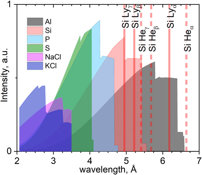
To estimate the emission spectra for elements with atomic numbers Z = 13–16, we used the atomic radiative-collision kinetic code PrismSPECT
![]()
Figure 1.Comparison of numerical photorecombination continuum emission spectra for a set of elements with atomic numbers
In the calculations, to enable a comparison between different materials and account for different electron configurations and ionization potentials, we simplified how to choose the conditions for our kinetic simulation by ensuring that the sum
| Element or compound | Z | ni × 1022 (ions/cm3) | ne × 1023 (electrons/сm3) | Te (eV) | Mean charge | PCE of H-like ion (Å) | PCE of He-like ion (Å) |
|---|---|---|---|---|---|---|---|
| Al | 13 | 6 | 6.9 | 430 | 11.5 | 5.61 | 6.18 |
| Si | 14 | 5 | 6.2 | 480 | 12.4 | 4.84 | 5.31 |
| P | 15 | 3.5 | 4.9 | 625 | 14 | 4.21 | 4.59 |
| S | 16 | 3.9 | 5.5 | 550 | 14.1 | 3.66 | 3.96 |
| NaCl | 2.2 | 3 | 1150 | 13.3 | 3.25 | 3.5 | |
| KCl | 1.6 | 2.7 | 1255 | 16.5 | 2.58 | 2.77 |
Table 1. Ion density ni, electron density ne, and electron temperature Te, which determine the emission spectra in
The results shown in
III. EXPERIMENTAL OPTIMIZATION OF THE CONFIGURATION OF THE X-RAY SOURCE TARGET
The possibility of using numerical spectra, such as those in
The experiment was conducted with the Vulcan petawatt laser at the Rutherford Appleton Laboratory, UK.
![]()
Figure 2.Top view of the experimental scheme. A solid target is irradiated by a p-polarized laser beam reflected from a focusing parabola and a plasma mirror. The front-side spectrometers are in the plane of the laser beam.
The following targets were used: (1) silicon foils with thicknesses from 0.5 μm to 30 µm, (2) aluminum foils with thicknesses from 5 μm to 25 µm, and (3) 2-µm silicon foils coated with 1.4 µm CH plastic on both front and rear sides. This plastic coating is transparent to the early part of the high-contrast laser pulse. The inertia of this coating helps prevent expansion of the buried layer until the arrival of the main laser pulse. This is discussed in Refs.
Three focusing spectrometers with spatial resolution (FSSRs)
![]()
Figure 3.Comparison of experimental data (grey curve) with a model spectrum (red curve). The experimental spectrum corresponds to 2-
The results in
The XRS emissivity or yield depends on the target configuration.
![]()
Figure 4.Dependences of experimentally measured conversion efficiencies on the thickness of solid foil targets: (a) emission from silicon targets integrated over the wavelength range 4.5 Å–5 Å and (b) emission from aluminum targets integrated over the range 5.15 Å–5.65 Å. The conversion efficiency is the ratio of the deposited laser energy and the energy of the emitted photons.
The data clearly reveal that there is an optimal target thickness of around 10 µm for both silicon and aluminum. For thinner targets, the decrease in emissivity is the result of a residual prepulse, which causes the target to expand. The decrease in plasma density reduces the recombination rate, so the X-ray yield drops, making thin targets unsuitable for XRS. When the target thickness was increased to 10 µm, the influence of the laser prepulse on the average plasma density reduced. A further increase in target thickness reduces the emissivity due to two factors. First, for a thicker target, the lowest electric potential is at the back surface of the target, which limits the refluxing of return currents of free electrons, reduces resistive heating, and reduces the target temperature. Second, the opacity of a thicker target reduces the emission from inner and rear layers.
The advantage of using plastic-coated targets to bury the emission layer is highlighted by the data point indicated by a black square in
IV. CONCLUSIONS
In this work, we discussed choosing a material and configuration of a solid target for use as a bright laser-plasma-based X-ray source. We show that low-Z materials, such as aluminum and silicon, can be used as X-ray backlighting in a hard X-ray range between 2 Å and 6 Å, which corresponds to photon energies of approximately 2 keV–6 keV. We focused on the photorecombination continuum emission of a solid-density plasma to create a featureless spectral continuum of high intensity, for use in, for example, XAS of WDM.
We studied experimentally spectrally resolved emissions from aluminum and silicon solid targets. We found that it is essential to use a high-contrast, high-intensity, and short-duration laser to create the X-ray sources and that an optimal target thickness of close to 10 µm is necessary. The lower emissivity of thinner targets, even with a high-contrast plasma pulse, is due to target expansion and a lowering of the average plasma density. A decrease of the emissivity for thicker targets is associated with a reduction of electron refluxing, resistive heating, and target opacity. A thin plastic coating on the target surface, which is transparent to the early part of a high-contrast laser pulse, helps to maintain the average plasma density and improve the X-ray emissivity.
The maximum number of the photons occurs for a 10-μm-thick Si foil and is about 1013 (assuming isotropic emission into 4π). However, when making XRS measurements during a specific high-power laser experiment, this value will likely decrease. The energy deposited into the main target usually exceeds that deposited into the XRS target. However, the plasma luminosity is directly proportional to the deposited laser energy. Therefore, the total main target luminosity is likely to be higher than the XRS one. Successful absorption spectroscopy seems to be impossible to perform in such conditions when recording photons emitted by both targets, as the signal-to-noise ratio is too high. However, most of the plasma emission comes from the hot laser–target interaction point and its surroundings, mainly as He-like characteristic emission lines. In a recent experimental campaign,
References
[1] F. B. Rosmej, O. Renner. Challenges of X-ray spectroscopy in investigations of matter under extreme conditions. Matter Radiat. Extremes, 4, 024201(2019).
[2] S. N. Ryazantsev, P. McKenna, I. Y. Skobelev, N. Booth, D. D. Arich, C. D. Murphy, T. A. Pikuz, P. S. Bratchenko, D. Farley, L. Doehl, K. L. Lancaster, S. B. Hansen, S. A. Pikuz, C. Spindloe, R. Kodama, C. D. Baird, P. Durey, A. Y. Faenov, J. Colgan, N. Woolsey. X-ray absorption spectroscopy study of energy transport in foil targets heated by petawatt laser pulses. Photonics Res., 6, 234(2018).
[3] I. Y. Skobelev, J. Fuchs, E. D. Filippov, A. Ciardi, S. A. Pikuz, G. Revet, B. Khiar, S. N. Chen, D. P. Higginson, D. Khaghani. X-ray spectroscopy evidence for plasma shell formation in experiments modeling accretion columns in young stars. Matter Radiat. Extremes, 4, 064402(2019).
[4] C. Bressler, M. Chergui. Ultrafast X-ray absorption spectroscopy. Chem. Rev., 104, 1781-1812(2004).
[5] J. P. Geindre, S. Bastiani-Ceccotti, S. Tzortzakis, J. C. Gauthier, S. Gary, P. Audebert, P. Renaudin, R. Shepherd, O. Peyrusse, C. Chenais-Popovics, V. Nagels, I. Matsushima, F. Girard. Time- and space-resolved X-ray absorption spectroscopy of aluminum irradiated by a subpicosecond high-power laser. J. Quant. Spectrosc. Radiat. Transfer, 99, 614(2006).
[6] S. Bastiani-Ceccotti, S. Gary, S. Tzortzakis, J.-P. Geindre, F. Girard, I. Matsushima, R. Shepherd, V. Nagels-Silvert, C. Chenais-Popovics, O. Peyrusse, J.-C. Gauthier, P. Renaudin, P. Audebert. Picosecond time-resolved X-ray absorption spectroscopy of ultrafast aluminum plasmas. Phys. Rev. Lett., 94, 025004(2005).
[7] J. D. Kilkenny, R. W. Eason, D. K. Bradley, G. N. Greaves. Improved laser-EXAFS studies of aluminium foil. J. Phys. C: Solid State Phys., 17, 5067-5074(1984).
[8] R. E. Rudd, B. A. Remington, J. S. Wark. From microjoules to megajoules and kilobars to gigabars: Probing matter at extreme states of deformation. Phys. Plasmas., 22, 090501(2015).
[9] P. A. Jaanimagi, V. A. Smalyuk, R. C. Mancini, S. P. Regan, J. A. Delettrez, P. B. Radha, R. Epstein, S. X. Hu, D. Li, H. Sawada, B. Yaakobi, T. R. Boehly, V. N. Goncharov, D. D. Meyerhofer, T. C. Sangster. Al 1s-2p absorption spectroscopy of shock-wave heating and compression in laser-driven planar foil. Phys. Plasmas, 16, 052702(2009).
[10] S. Toleikis, J. Gaudin, H. J. Lee, G. Morard, E. Galtier, A. Denoeud, C. Fourment, A. Ravasio, F. Dorchies, A. Benuzzi-Mounaix, T. Vinci, F. Guyot, R. Musella, K. Miyanishi, R. Kodama, Y. Feng, B. Nagler, S. Mazevet, D. Zhu, M. Nakatsutsumi, U. Zastrau, M. Koenig, J. Bouchet, M. Harmand, V. Recoules, N. Ozaki. X-ray absorption spectroscopy of iron at multimegabar pressures in laser shock experiments. Phys. Rev. B., 92, 024108(2015).
[11] Y. Ping, D. E. Fratanduono, R. F. Smith, J. R. Rygg, D. C. Swift, G. W. Collins, B. Yaakobi, F. Coppari, T. R. Boehly, D. G. Braun, J. H. Eggert, S. Hamel, D. G. Hicks. Solid iron compressed up to 560 GPa. Phys. Rev. Lett., 111, 065501(2013).
[12] T. J. B. Collins, H. E. Lorenzana, P. G. Allen, B. Yaakobi, D. D. Meyerhofer, S. M. Pollaine, T. R. Boehly, B. A. Remington, J. H. Eggert. EXAFS measurement of iron bcc-to-hcp phase transformation in nanosecond-laser shocks. Phys. Rev. Lett., 95, 075501(2005).
[13] C. L. Jackson, A. Djaoui, A. Cole, B. Shiwai, R. W. Eason, T. A. Hall, S. L. Rose, P. Apte. Experimental observation of ion correlation in a dense laser-produced plasma. Phys. Rev. Lett., 60, 2034-2037(1988).
[14] V. Jonauskas, H.-K. Chung, C. Ramsbottom, F. P. Keenan, D. A. Liedahl, W. H. Goldstein, S. J. Rose, P. T. Springer, M. E. Foord, J. E. Bailey, K. B. Fournier, M. E. Cuneo, R. F. Heeter, R. Kisielius, P. A. M. van Hoof. Study of X-ray photoionized Fe plasma and comparisons with astrophysical modeling codes. J. Quant. Spectrosc. Radiat. Transfer, 99, 712-729(2006).
[15] D. Jobe, P. Woodruff, R. B. Spielman, C. Deeney, J. E. Bailey, G. A. Chandler, J. J. MacFarlane, M. R. Douglas, P. Lake, P. Wang, T. J. Nash, D. S. Nielsen. X-ray absorption spectroscopy measurements of thin foil heating by Z -pinch radiation. Phys. Rev. E., 66, 046416(2002).
[16] G. Xiong, S. Liu, S. Jiang, J. Yang, G. Yang, Z. Yuan, Y. Zhao, B. Zhang, H. Li, Y. Ding, J. Zhang, H. Zhang, X. Meng, Y. Xu, Z. Zheng, J. Yan. L- and M-shell absorption measurements of radiatively heated Fe plasma. Phys. Plasmas., 19, 113302(2012).
[17] K. Falk. Experimental methods for warm dense matter research. High Power Laser Sci. Eng., 6, e59(2018).
[18] F. Dorchies, S. Fourmaux, P. Audebert, L. Lancia, S. Mazevet, C. Fourment, O. Peyrusse, J. J. Santos, P. Antici, J. Fuchs, M. Harmand, S. Hulin, P. Renaudin, A. Mancic, A. Lévy, V. Recoules, M. Nakatsutsumi. X-ray absorption for the study of warm dense matter. Plasma Phys. Control. Fusion., 51, 124021(2009).
[19] H. J. Lee, R. W. Falcone, P. A. Ni, C. P. Weber, J. Feng, A. J. Nelson, P. A. Heimann, D. Prendergast, A. A. Correa, Y. Ping, K. Engelhorn, R. W. Lee, T. Ogitsu, B. I. Cho. Electronic structure of warm dense copper studied by ultrafast x-ray absorption spectroscopy. Phys. Rev. Lett., 106, 167601(2011).
[20] M. Störmer, J. Bouchet, M. Nakatsutsumi, K. Engelhorn, C. Ozkan, C. Fourment, M. Harmand, P. A. Heimann, H. J. Lee, E. Galtier, S. Toleikis, F. Dorchies, B. Nagler, T. Tschentscher, J. Gaudin, P. M. Leguay, B. I. Cho, V. Recoules. Time evolution of electron structure in femtosecond heated warm dense molybdenum. Phys. Rev. B., 92, 144201(2015).
[21] O. A. Hurricane, R. Betti. Inertial-confinement fusion with lasers. Nat. Phys., 12, 435-448(2016).
[22] A. Casner, S. Baton, D. Batani, X. Ribeyre, M. Koenig, C. Rousseaux, W. Theobald, M. Hohenberger, O. Klimo, C. Labaune, S. Depierreux, G. Schurtz, V. T. Tikhonchuk. Physics issues for shock ignition. Nucl. Fusion., 54, 054009(2014).
[23] S. B. Hansen, J.-C. Pain, B. G. Wilson, G. A. Rochau, F. Gilleron, C. A. Iglesias, G. P. Loisel, R. C. Mancini, C. Blancard, S. N. Nahar, P. Cosse, C. J. Fontes, T. Nagayama, J. J. MacFarlane, J. E. Bailey, A. K. Pradhan, G. Faussurier, D. P. Kilcrease, C. Orban, I. Golovkin, M. Sherrill, J. Colgan. A higher-than-predicted measurement of iron opacity at solar interior temperatures. Nature, 517, 56-59(2015).
[24] S. N. Nahar, A. K. Pradhan. Large enhancement in high-energy photoionization of Fe XVII and missing continuum plasma opacity. Phys. Rev. Lett., 116, 235003(2016).
[25] G. C. O’Neil, K. L. Silverman, J. Uhlig, M. L. Dowell, C. L. Cromer, J. N. Ullom, L. Miaja-Avila, R. Jimenez, A. S. Hoover. Laser plasma x-ray source for ultrafast time-resolved x-ray absorption spectroscopy. Struct. Dyn., 2, 024301(2015).
[26] P. McKenna, P. Durey, C. D. Murphy, M. V. Sedov, A. Y. Faenov, C. D. Baird, K. Y. Platonov, N. Woolsey, A. A. Andreev, N. Booth, T. A. Pikuz, I. Y. Skobelev, S. N. Ryazantsev, D. Farley, C. Spindloe, S. A. Pikuz, L. Doehl, K. L. Lancaster, R. Kodama. Features of the generation of fast particles from microstructured targets irradiated by high intensity, picosecond laser pulses. Laser Part. Beams, 37, 176-183(2019).
[27] A. S. Pirozhkov, H. Kiriyama, K. Kondo, K. Kondo, H. Sakaki, M. A. Alkhimova, A. Sagisaka, Y. Fukuda, S. A. Pikuz, T. A. Pikuz, A. Y. Faenov, K. Nishitani, M. Kando, T. Miyahara, R. Kodama, M. Nishiuchi, I. Y. Skobelev, K. Ogura, N. P. Dover, Y. Watanabe. High resolution X-ray spectra of stainless steel foils irradiated by femtosecond laser pulses with ultra-relativistic intensities. Opt. Express., 25, 29501(2017).
[28] A. G. Es’kov, N. M. Umrikhin, I. M. Poznyak, V. V. Gavrilov, S. A. Pikuz, D. A. Toporkov, I. Y. Skobelev, D. M. Kochnev, S. N. Ryazantsev, A. M. Zhitlukhin. High-power X-ray line radiation of the plasma produced in a collision of high-energy plasma flows. Plasma Phys. Rep., 44, 820(2018).
[29] J. J. Santos, S. Hulin, D. Descamps, A. Lévy, C. Goyon, S. Petit, O. Peyrusse, F. Dorchies, P. Combis, M. Harmand, P. M. Leguay, C. Fourment. Unraveling the solid-liquid-vapor phase transition dynamics at the atomic level with ultrafast x-ray absorption near-edge spectroscopy. Phys. Rev. Lett., 107, 245006(2011).
[30] V. Recoules, F. Dorchies. Non-equilibrium solid-to-plasma transition dynamics using XANES diagnostic. Phys. Rep., 657, 1-26(2016).
[31] S. Fourmaux, M. Harmand, L. Lecherbourg, J. C. Kieffer, M. Servol. High repetition rate laser produced soft x-ray source for ultrafast x-ray absorption near edge structure measurements. Rev. Sci. Instrum., 78, 113104(2007).
[32] D. A. Hammer, T. Shelkovenko, A. D. Cahill, S. A. Pikuz, C. L. Hoyt. A doubly curved elliptical crystal spectrometer for the study of localized x-ray absorption in hot plasmas. Rev. Sci. Instrum., 85, 103114(2014).
[33] D. A. Hammer, S. A. Pikuz, T. A. Shelkovenko. A review of projection radiography of plasma and biological objects in X-Pinch radiation. Plasma Phys. Rep., 42, 226-268(2016).
[34] M. Beye, M. Burkhardt, J. Stöhr, Y. Acremann, A. Föhlisch, T. Beeck, F. Sorgenfrei, W. Wurth, A. Scherz, W. F. Schlotter, D. P. Bernstein, A. Pietzsch. Near edge x-ray absorption fine structure spectroscopy with x-ray free-electron lasers. Appl. Phys. Lett., 95, 134102(2009).
[35] M. Hansson, O. Lundh, I. Gallardo González, K. Falk, J. C. Wood, S. P. D. Mangles, A. Persson, J. Björklund Svensson, H. Ekerfelt, M. Šmíd. Highly efficient angularly resolving x-ray spectrometer optimized for absorption measurements with collimated sources. Rev. Sci. Instrum., 88, 063102(2017).
[36] A. Lifschitz, K. Ta Phuoc, L. Lecherbourg, J.-P. Goddet, P. Renaudin, B. Mahieu, F. Dorchies, N. Jourdain. Probing warm dense matter using femtosecond X-ray absorption spectroscopy with a laser-produced betatron source. Nat. Commun., 9, 3276(2018).
[37] R. A. Baggott, K. Falk, S. J. Rose, E. Gerstmayr, S. Dann, O. Lundh, N. Bourgeois, S. P. D. Mangles, B. Kettle, R. Watt, M. Šmíd, C. Spindloe, N. Lemos, A. G. R. Thomas, M. J. V. Streeter, A. E. Hussein, F. Albert, Y. Ma, J. M. Cole, I. Gallardo González, N. C. Lopes, D. R. Symes. Single-shot multi-keV x-ray absorption spectroscopy using an ultrashort laser-wakefield accelerator source. Phys. Rev. Lett., 123, 254801(2019).
[38] B. M. Hegelich, D. Papp, S. Q. Wu, H. Chen, R. Tommasini, A. L. Milder, F. V. Hartemann, F. Albert, J. Moody, B. B. Pollock, W. Schumaker, C. Joshi, J. E. Ralph, K. A. Marsh, J. L. Shaw, C. Goyon, A. Pak, A. M. Saunders, G. J. Williams, P. King, S. H. Glenzer, J. Park, P. Michel, N. Lemos, R. Polanek. X-ray sources using a picosecond laser driven plasma accelerator. Phys. Plasmas., 26, 083110(2019).
[39] S. Fourmaux, M. Z. Mo, A. Saraf, Y. Tsui, Z. Chen, R. Masoud, R. Fedosejevs, K. Otani, A. Ng, S. Kerr, J.-C. Kieffer. Measurements of ionization states in warm dense aluminum with betatron radiation. Phys. Rev. E., 95, 053208(2017).
[40] A. Jonas, R. Jung, B. Kanngießer, I. Mantouvalou, K. Witte, H. Stiel. Optimizing soft X-ray NEXAFS spectroscopy in the laboratory. Proc. SPIE, 10243, 1024308(2017).
[41] T. Blenski, V. Silvert, F. Thais, G. Loisel, P. Arnault, J. Fariaut, T. Caillaud, S. Turck-Chièze, B. Villette, J.-C. Pain, S. Bastiani-Ceccotti, M. Poirier, C. Reverdin, F. Gilleron, W. Fölsner. Absorption spectroscopy of mid and neighboring Z plasmas: Iron, nickel,copper and germanium. High Energy Density Phys., 5, 173-181(2009).
[42] B. E. Campbell, P. J. Mallozzi, H. M. Epstein, R. E. Schwerzel. Laser-EXAFS: Fast extended x-ray absorption fine structure spectroscopy with a single pulse of laser-produced x-rays. Science, 206, 353-355(1979).
[43] A. Bartnik, T. Fok, K. A. Janulewicz, H. Fiedorowicz, P. Wachulak. EXAFS of titanium LIII edge using a compact laboratory system based on a laser-plasma soft X-ray source. Appl. Phys. B., 126, 11(2020).
[44] B. A. Remington, E. M. Garcia, D. B. Thorn, H.-S. Park, M. B. Schneider, R. S. Craxton, F. Coppari, A. Krygier, Y. Ping, G. E. Kemp, J. H. Eggert, J. M. McNaney. Developing a high-flux, high-energy continuum backlighter for extended x-ray absorption fine structure measurements at the National Ignition Facility. Rev. Sci. Instrum., 89, 10F114(2018).
[45] Z. Wang, Y. Hu, S. Jiang, J. Zhang, Q. Xue, Q. Ye. X-ray source improvements for EXAFS measurement on SGIII prototype facility. AIP Adv., 10, 055313(2020).
[46] D. D. Meyerhofer, B. Yaakobi, R. P. J. Town, F. J. Marshall, T. R. Boehly. Extended x-ray absorption fine-structure experiments with a laser-imploded target as a radiation source. J. Opt. Soc. Am. B., 20, 238(2003).
[47] Y. J. Son, J. H. Sung, Y. H. Jang, C. W. Lee, J. W. Yoon, S. K. Lee, J. M. Yang, J. Y. Yoo, H. W. Lee, C. H. Nam. 42 PW, 20 fs Ti:sapphire laser at 01 Hz. Opt. Lett., 42, 2058(2017).
[48] I. Dancus, M. Zeng, L. D’Alessi, D. Filipescu, N. V. Zamfir, S. Gales, D. G. Ghita, I. Andrei, F. Negoita, C. Matei, P. Ghenuche, K. A. Tanaka, K. Seto, D. L. Balabanski, D. Stutman, M. O. Cernaianu, S. Ataman, B. Diaconescu, D. Ursescu, C. A. Ur, O. Tesileanu, N. Djourelov. The extreme light infrastructure—Nuclear physics (ELI-NP) facility: New horizons in physics with 10 PW ultra-intense lasers and 20 MeV brilliant gamma beams. Rep. Prog. Phys., 81, 094301(2018).
[49] F. Mathieu, K. Genevrier, L. Ranc, J. P. Zou, N. Lebas, C. Le Blanc, P. Georges, L. Martin, A. Pellegrina, D. N. Papadopoulos, P. Monot, F. Druon, P. Ramirez, P. Audebert. High-contrast 10 fs OPCPA-based front end for multi-PW laser chains. Opt. Lett., 42, 3530(2017).
[50] W. Li, S. Li, L. Yu, Y. Li, Y. Xu, Y. Liu, Z. Xu, X. Lu, Y. Leng, Y. Yang, F. Wu, C. Wang, X. Yang, Z. Liu, R. Li. High-contrast front end based on cascaded XPWG and femtosecond OPA for 10-PW-level Ti:sapphire laser. Opt. Express., 26, 2625(2018).
[51] Y. Miyasaka, A. S. Pirozhkov, M. Mori, T. Z. Esirkepov, J. K. Koga, K. Kondo, K. Kondo, A. Sagisaka, N. P. Dover, M. Nishiuchi, H. Sakaki, M. Kando, K. Ogura, Y. Fukuda, H. Kiriyama. High-contrast high-intensity repetitive petawatt laser. Opt. Lett., 43, 2595(2018).
[52] R. J. Clarke, C. J. Hooker, F. Y. Khattak, J. J. Angulo-Gareta, A. J. Langley, P. S. Foster, D. Neely, M. J. Lamb, E. J. Divall, D. Riley. Kα yields from Ti foils irradiated with ultrashort laser pulses. Phys. Rev. E., 71, 016406(2005).
[53] G. Hays, K. B. Wharton, J. Zweiback, T. Ditmire, C. D. Boley, A. M. Komashko, T. E. Cowan, J. Crane, A. M. Rubenchik. Effects of nonionizing prepulses in high-intensity laser-solid interactions. Phys. Rev. E., 64, 025401(2001).
[54] P. Neumayer, A. L. Kritcher, S. H. Glenzer, C. Niemann, H. Robey, M. K. Urry, O. L. Landen, E. Morse. K-alpha conversion efficiency measurements for X-ray scattering in inertial confinement fusion plasmas. High Energy Density Phys., 3, 156-162(2007).
[55] G. Gregori, S. C. Wilks, S. N. Chen, P. K. Patel, F. N. Beg, D. Riley, R. B. Stephens, J. A. King, S. B. Hansen, J. R. Pasley, R. L. Weber, S. H. Glenzer, F. Y. Khattak, H.-K. Chung, A. J. Mackinnon, R. R. Freeman, E. Garcia Saiz, M. M. Notley, R. G. Evans. Creation of hot dense matter in short-pulse laser-plasma interaction with tamped titanium foils. Phys. Plasmas., 14, 102701(2007).
[56] I. Y. Skobelev, A. S. Martynenko, C. Baird, S. N. Ryazantsev, S. A. Pikuz, C. D. Murphy, N. Woolsey, N. Booth, P. Durey, R. Kodama, P. McKenna, L. Doehl, T. A. Pikuz, D. Farley, K. Lancaster, A. Y. Faenov, C. Spindloe. Effect of plastic coating on the density of plasma formed in Si foil targets irradiated by ultra-high-contrast relativistic laser pulses. Phys. Rev. E., 101, 043208(2020).
[57] B. V. Oliver, R. B. Campbell, J. J. MacFarlane, T. A. Mehlhorn, P. R. Woodruff, D. R. Welch, I. E. Golovkin. Simulation of the ionization dynamics of aluminum irradiated by intense short-pulse lasers, 457(2004).
[58] P. Wang, J. J. MacFarlane, N. A. Pereyra, I. E. Golovkin, P. R. Woodruff. SPECT3D—A multi-dimensional collisional-radiative code for generating diagnostic signatures based on hydrodynamics and PIC simulation output. High Energy Density Phys., 3, 181(2007).
[59] H. K. Chung, M. G. Brookes, M. P. Hill, J. Dunn, R. W. Lee, L. M. R. Hobbs, D. J. Hoarty, J. Morton, H. Chen, P. Beiersdorfer, C. R. D. Brown, S. F. James, J. W. O. Harris, E. Von Marley, R. Shepherd, G. Brown, P. Allan, J. Emig. Observations of the effect of ionization-potential depression in hot dense plasma. Phys. Rev. Lett., 110, 265003(2013).
[60] B. I. Cho, U. Zastrau, T. R. Preston, J. J. Turner, D. S. Rackstraw, T. Burian, G. L. Dakovski, J. Chalupský, S. M. Vinko, R. W. Lee, P. Heimann, L. Juha, H. K. Chung, M. Holmes, B. Barbrel, V. Hájková, O. Ciricosta, K. Engelhorn, J. Krzywinski, J. S. Wark, S. Toleikis. Measurements of continuum lowering in solid-density plasmas created from elements and compounds. Nat. Commun., 7, 11713(2016).
[61] D. Mihalas, D. G. Hummer. The equation of state for stellar envelopes. I. An occupation probability formalism for the truncation of internal partition functions. Astrophys. J., 331, 794(1988).
[62] C. Hernandez-Gomez, P. Holligan, P. A. Norreys, J. L. Collier, B. Fell, R. J. Clarke, D. A. Pepler, W. J. Lester, M. H. R. Hutchinson, A. J. Frackiewicz, D. Neely, A. Kidd, R. W. W. Wyatt, P. A. Brummitt, C. J. Reason, C. N. Danson, D. R. Neville, T. B. Winstone, W. Shaikh, S. Hancock, S. Hawkes, B. E. Wyborn, I. O. Musgrave. Vulcan petawatt—An ultra-high-intensity interaction facility. Nucl. Fusion, 44, S239-S246(2004).
[63] S. V. Bulanov, T. Tajima, G. A. Mourou. Optics in the relativistic regime. Rev. Mod. Phys., 78, 309-371(2006).
[64] J. C. Gauthier, P. Audebert, F. Quéré, O. Gobert, G. Doumy, P. Martin, M. Perdrix, T. Wittmann, J.-P. Geindre. Complete characterization of a plasma mirror for the production of high-contrast ultraintense laser pulses. Phys. Rev. E., 69, 026402(2004).
[65] F. Krausz, B. Dromey, S. Kar, D. Neely, M. Zepf, D. Adams, R. Hörlein, G. D. Tsakiris, Y. Nomura, P. Foster, K. Markey. High contrast plasma mirror: Spatial filtering and second harmonic generation at 1019 W cm−2. New J. Phys., 10, 083002(2008).
[66] C. Hernandez-Gomez. Overview of the central laser facility (CLF), 6-8(2017).
[67] T. A. Shelkovenko, A. I. Erko, G. V. Ivanenkov, T. A. Pikuz, V. M. Dyakin, A. R. Mingaleev, A. Y. Faenov, S. A. Pikuz, B. A. Bryunetkin, V. M. Romanova. High-performance x-ray spectroscopic devices for plasma microsources investigations. Phys. Scr., 50, 333-338(1994).
[68] D. A. Arich, S. A. Pikuz, I. Y. Skobelev, T. A. Pikuz, A. Y. Faenov, M. A. Alkhimova. Accounting for the instrument function of crystal spectrometers operating in many reflection orders in the diagnostics of laser plasma from its continuum spectrum. Quantum Electron., 48, 749-754(2018).
[69] G. Boutoux, T. Sakaki, J. J. Honrubia, O. N. Rosmej, A. Sauteray, J. J. Santos, D. Khaghani, A. Franz, L. Giuffrida, S. Pikuz, L. Antonelli, A. Schönlein, D. Batani, J. Jacoby, P. Neumayer, A. Debayle. Generation and characterization of warm dense matter isochorically heated by laser-induced relativistic electrons in a wire target. EPL (Europhys. Lett.), 114, 45002(2016).

Set citation alerts for the article
Please enter your email address
© Copyright 2018-2021 | Chinese Laser Press. All Rights Reserved 沪ICP备15018463号-20