
Tianchen Zhao, Qiang Ma, Yajie Bian, Yuyi Zhang, Yiting Liu, Xiaolei Zhang, Botao Wu, E Wu, Shitao Lou, Qingyuan Jin, "Strong coupling with absorption and emission features of Ag@Au hollow nanoshells interacting with J-aggregated dye molecules," Chin. Opt. Lett. 19, 123602 (2021)
- Chinese Optics Letters
- Vol. 19, Issue 12, 123602 (2021)
Abstract
1. Introduction
Localized surface plasmon resonance (LSPR) is the collective resonance of the surface electrons of metal nanoparticles (NPs) when interacting with the electromagnetic field of incident light[
J-aggregates are the assemblies of molecules with a very intense, narrow absorption band (J band), which is red-shifted to a longer wavelength by comparison with the monomer absorption band[
In recent years, strong coupling systems based on metal nanostructures and J-aggregate systems have increasingly become a research hotspot in the field of nanoscience, including several research areas, such as the research on LSPR-based biosensors[
Sign up for Chinese Optics Letters TOC. Get the latest issue of Chinese Optics Letters delivered right to you!Sign up now
In this paper, we choose the fairly stable J-aggregated TDBC molecules and the triangular Ag nanoprisms or Ag@Au HNS with excellent optical and structural properties to build the tunable plexcitons strong coupling system. In our experiments, Rabi splitting can be clearly observed in pure Ag NPs or Ag@Au HNS with J-aggregated dyes at RT, with the changing of the split peaks’ intensities and positions due to the different LSPR modes of the two kinds of NPs. Moreover, the photoluminescence (PL) intensity tends to decrease with the increasing temperature, showing more prominent local field enhancement effects in the lower temperature. It indicates weaker coupling interaction between the metallic NPs and the dyes in the higher temperature, to a certain extent, identical with the results of temperature-dependent observations in the similar plexcitons coupling system reported previously[
2. Experimental Section
2.1. Sample preparation
2.1.1. Ag nanoprisms and Ag@Au HNS synthesis
The preparation of Ag nanoprisms is divided into seed preparation and particle synthesis. First, aqueous trisodium citrate (5 mL, 2.5 mmol/L), aqueous poly (sodium styrenesulphonate) (PSSS; 0.25 mL, 500 mg/L), and freshly prepared aqueous
The Ag@Au HNS synthesis was also completed in two steps. In the first step, 9 mL of Ag NPs solution were refluxed for 10 min. In the second step, the
2.1.2. Ag nanoprisms or Ag@Au HNS/J-aggregate hybrid fabrication
TDBC sodium salt was purchased from Shanghai Rechem science Co., Ltd. Ag@Au HNS/TDBC hybrid was produced by combining TDBC aqueous solution (2200 µL, 100 µmol/L) and 5 mL Ag@Au HNS solution under gentle magnetic stirring. In the same way, Ag NPs/TDBC hybrid was produced by combining TDBC aqueous solution (2500 µL, 100 µmol/L) and 6 mL Ag NPs solution under gentle magnetic stirring. After at least 24 h, both hybrid syntheses were centrifuged with water (10,000 r/min, 7 min) to remove excess TDBC dye molecules.
2.1.3. APTES functionalization
All of the samples were prepared in aqueous solution. Nevertheless, fluorescent spectra at low temperatures require them to be assembled on solid substrates. Now, we will explain how to assemble the hybrid nanostructures on a glass substrate[
2.2. Characterization
A PERSEE Tu-1901 instrument was used for the absorption spectral test. A liquid nitrogen cooled charge coupled device (CCD) with a spectrometer (Princeton Instruments) was used for the steady-state PL spectral detection. The PL was excited by a continuous light source (mercury lamp, setting the illumination wavelength at 532 nm). A microchannel plate photomultiplier tube (Hamamatsu) was used for photon counting. Transmission electron microscope (TEM) analysis was performed using a TECNAI G2F20 instrument, and scanning TEM (STEM) analysis was performed using a JEM2100F instrument; the samples were prepared by drop-casting 10 µL solution onto a Lacey Formvar/Carbon Film Coated Grids overnight and allowing the solvent (water) to evaporate naturally.
2.3. Simulations
The commercial software FDTD Solutions from Lumerical Solution, Inc., Canada, which is based on the finite-difference time-domain method, was used to simulate plexcitons coupling. A total field-scattered field source is injected onto Ag NPs or Ag@Au HNS without and with a TDBC shell, and the simulation wavelength ranges from 400 to 800 nm. The measured dielectric constants of Ag and Au are used in the simulation, which are included in Refs. [22,23], respectively. The optical properties of TDBC are described by a Lorentzian function,
3. Results and Discussion
3.1. Experimental strong coupling of plasmons and excitons
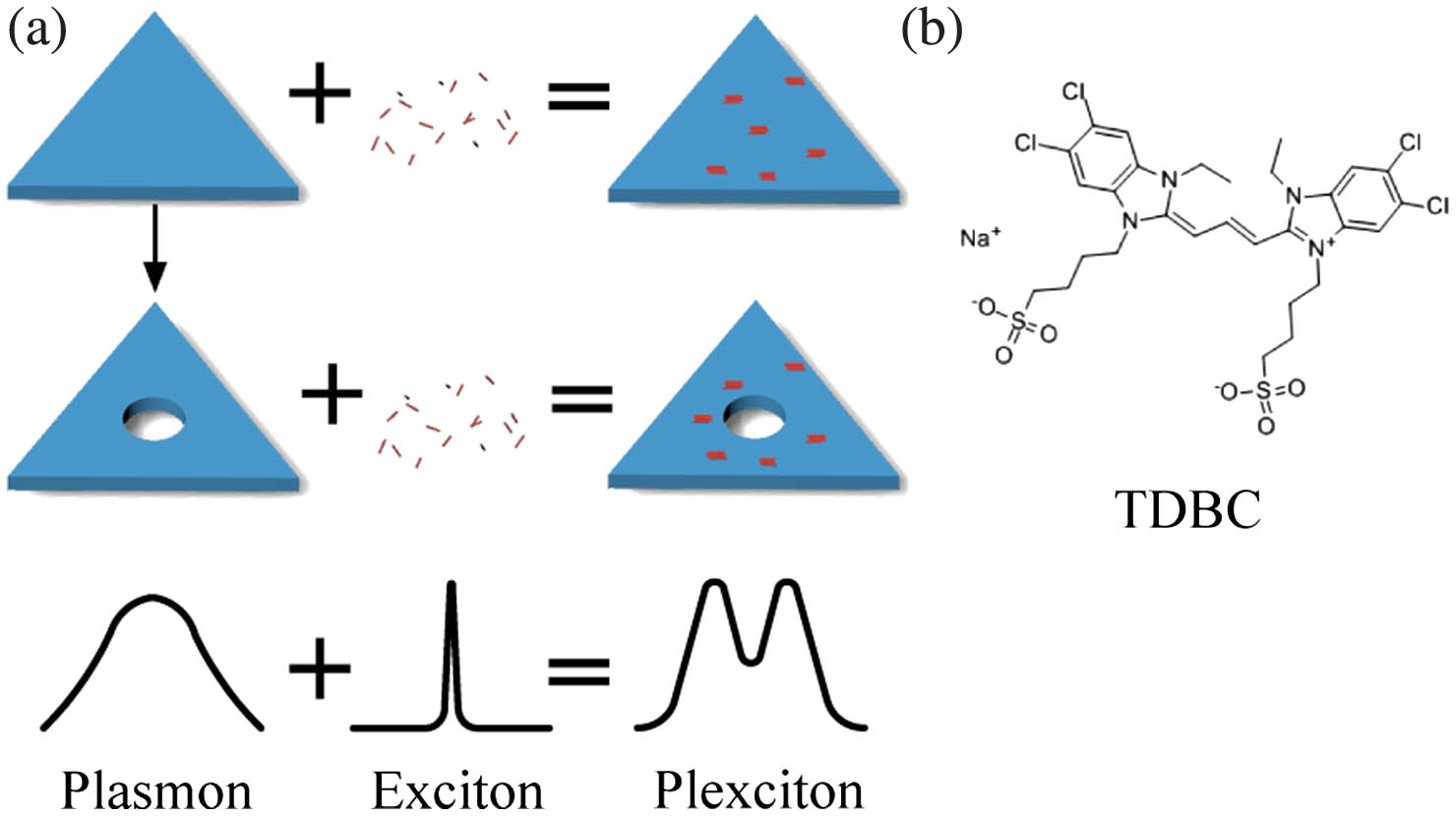
The self-assembly processes of J-aggregated dyes in Ag nanoprisms and Ag@Au HNS are shown in Fig. 1(a). Figure 1(b) shows the chemical structure of a TDBC molecule. In the mixture of the NP colloidal solution and the TDBC solution, the citrate is adsorbed to the Au NPs surface through oxygen and can be easily replaced by TDBC molecules. So, the TDBC self-assembly is realized through ligand exchange reaction between the citrate and TDBC molecules[
![]()
Figure 1.(a) Schematic diagram of the hybrid of Ag nanoprisms and Ag@Au HNS embedded in an ensemble of J-aggregate (TDBC). (b) The structural formula of a TDBC molecule.
The triangular edge structure of Ag nanoprisms and the Ag@Au HNS can be seen from the TEM images in Figs. 2(a) and 2(b) and the STEM images in Figs. 2(f) and 2(h). At first, Ag nano-triangular prisms were synthesized as the templates. When Au ions were added, the Ag atoms were replaced by electrochemical reduction (galvanic replacement reaction, GRR)[
| Element | Weight (%) | Atomic Fraction (%) |
|---|---|---|
| Ag | 73.56 | 83.55 |
| Au | 26.44 | 16.45 |
Table 1. EDX Spectrum Analysis of Ag@Au Hollow Nanostructure
![]()
Figure 2.TEM images of (a) Ag NPs, (b) Ag@Au HNS, (c) Ag NPs/TDBC, and (d) Ag@Au HNS/TDBC. (e) and (g) Mapping of elements (distribution of Ag/Au element) of Ag NPs and Ag@Au HNS. (f) and (h) STEM images of Ag NPs and Ag@Au HNS. (i) The energy dispersive X-ray spectroscopy (EDX) analysis of Ag@Au HNS.
![]()
Figure 3.(a) Ultraviolet-visible absorption spectrum of TDBC dye molecules. (b) Ultraviolet-visible absorption spectra of four nanostructures (black line indicates Ag NPs, red line indicates Ag@Au HNS, blue line indicates Ag NPs/TDBC, and green line indicates Ag@Au HNS/TDBC).
Figure 3(a) shows the absorption spectrum of TDBC dye molecules. The J-aggregate absorption peak is around 587 nm, which is consistent with the literatures[
For the LSPR of Ag@Au HNS [586 nm, red line in Fig. 3(b)], whose mode matches the exciton peak of TDBC (587 nm), the strongly coupled hybrid exhibits obvious mode splitting. The absorption spectrum of the corresponding system (Ag@Au HNS/TDBC) splits into two new polariton peaks at 551 nm (2251 meV) and 647 nm (1917 meV), which exhibit comparable intensities. The Rabi splitting is around 334 meV, as shown in the green line in Fig. 3(b), which is smaller than that of Ag NPs/TDBC. Compared with solid core Ag nanoparticles, Ag@Au HNS has a unique voluminous void space, so it can improve radiation absorption efficiencies through “light trapping” effects. Simultaneously, the hollow materials can enhance light harvesting[
Although the Rabi splitting of the absorption peak already indicates strong coupling between Ag NPs/Ag@Au HNS and TDBC, in order to better understand the physical mechanism involved, we adopt a more general strong coupling criterion[
The fluorescent spectra of the hybrid system of Ag NPs/TDBC and Ag@Au HNS/TDBC at RT are shown in Fig. 4(a). The fluorescence peaks of the two systems are both around 577 nm. Figures 4(b) and 4(c) show the variation curves of the intensity of the hybrid system of Ag NPs/TDBC and Ag@Au HNS/TDBC when the temperature gradually decreases from RT (300 K) to 100 K. In Fig. 4(b), the fluorescence intensity of the hybrid system of Ag NPs/TDBC shows an upward trend as the temperature decreases. At certain temperatures (240 K, 180 K, and 100 K), there will be a downward trend, but the overall trend remains rising. In Fig. 4(c), the fluorescence intensity of the hybrid system of Ag@Au HNS/TDBC also shows an upward trend with the decrease of temperature. At a certain temperature (only 100 K), there will be a downward trend. But, the overall trend keeps rising. It can be seen that when the temperature gradually decreases, there appears to be more local field enhancement. Up to a certain extent, the results illustrate that the coupling between Ag NPs/Ag@Au HNS and TDBC is strongly affected by temperature changes, similar to literature information[
![]()
Figure 4.(a) Fluorescent spectra of Ag NPs/TDBC and Ag@Au HNS/TDBC. (b), (c) Temperature-dependent fluorescence intensity curves of Ag NPs/TDBC and Ag@Au HNS/TDBC measured at T = 100–300 K.
3.2. Simulated strong coupling of plasmons and excitons
To explain the observed positions and intensities of the absorption peaks varying with the LSPR modes and the structures of the Ag NPs or Ag@Au HNS with J-aggregated dyes, the near field distributions for the two systems were simulated using the FDTD method. As shown in Figs. 5(a)–5(h), strong electric fields (
![]()
Figure 5.Electric field (|E/E0|) distribution images for the (a) pure Ag individual NP with LSPR peak at 565 nm, (b) TDBC-doped Ag NPs ensemble with LSPR peak at the upper branch (ω+) maximum (544 nm), (c) TDBC-doped Ag NPs ensemble with LSPR peak at the minimum (577 nm), (d) J-aggregate-doped Ag NPs ensemble with LSPR peak at the lower branch (ω−) maximum (607 nm), (e) Ag@Au HNS individual nanoparticle with LSPR peak at 589 nm, (f) TDBC-doped Ag@Au HNS ensemble with LSPR peak at the upper branch (ω+) maximum (559 nm), (g) TDBC-doped Ag@Au HNS ensemble with LSPR peak at the minimum (583 nm), and (h) J-aggregate-doped Ag@Au HNS ensemble with LSPR peak at the lower branch (ω−) maximum (612 nm). (i) Simulation extinction spectra of four nanostructures (black line indicates Ag NPs, red line indicates Ag@Au HNS, blue line indicates Ag NPs/TDBC, and green line indicates Ag@Au HNS/TDBC).
4. Conclusion
In summary, bimetallic HNSs with controlled LSPR were synthesized. After TDBC molecules were deposited on the bimetallic HNS, the absorption spectra and PL spectra were studied. Strong coupling was observed by precisely adjusting the LSPR of NPs to match the molecular absorption band; meanwhile, the coupling phenomenon is stronger than that of Ag nanoprisms. In the variable temperature experiment, the coupling strength can be inferred by observing the change of PL intensity, which tends to decrease with the temperature increasing. Finally, the strong coupling experimental results have been successfully verified by the FDTD simulation.
References
[1] L. J. Sherry, R. C. Jin, C. A. Mirkin, G. C. Schatz, R. P. Van Duyne. Localized surface plasmon resonance spectroscopy of single silver triangular nanoprisms. Nano Lett., 6, 2060(2006).
[2] Q. Zhang, J. J. Ren, X. K. Duan, H. Hao, Q. H. Gong, Y. Gu. Enhancing coupling coefficient in a hybrid nanotoroid-nanowire system. Chin. Opt. Lett., 17, 032702(2019).
[3] X. R. Ma, H. Cheng, J. W. Hou, G. H. Wu, X. Y. Lü, X. X. Zheng, C. Chen. Detection of breast cancer based on novel porous silicon Bragg reflector surface-enhanced Raman spectroscopy-active structure. Chin. Opt. Lett., 18, 051701(2020).
[4] L. C. Sun, Z. Li, J. S. He, P. J. Wang. Strong coupling with directional absorption features of Ag@Au hollow nanoshell/J-aggregate heterostructures. Nanophotonics, 8, 1835(2019).
[5] D. G. Baranov, M. Wersall, J. Cuadra, T. J. Antosiewicz, T. Shegai. Novel nanostructures and materials for strong light–matter interactions. ACS Photonics, 5, 24(2018).
[6] Y. K. Tang, X. T. Yu, H. F. Pan, J. Q. Chen, B. Audit, F. Argoul, S. J. Zhang, J. H. Xu. Numerical study of novel ratiometric sensors based on plasmon–exciton coupling. Appl. Spectrosc., 71, 2377(2017).
[7] G. Zengin, M. Wersäll, S. Nilsson, T. J. Antosiewicz, M. Käll, T. Shegai. Realizing strong light–matter interactions between single-nanoparticle plasmons and molecular excitons at ambient conditions. Phys. Rev. Lett., 114, 157401(2015).
[8] G. Zengin, G. Johansson, P. Johansson, T. J. Antosiewicz, M. Käll, T. Shegai. Approaching the strong coupling limit in single plasmonic nanorods interacting with J-aggregates. Sci. Rep., 3, 3074(2013).
[9] D. D. Lekeufack, A. Brioude, A. W. Coleman, P. Miele, J. Bellessa, L. D. Zeng, P. Stadelmann. Core-shell gold J-aggregate nanoparticles for highly efficient strong coupling applications. Appl. Phys. Lett., 96, 253107(2010).
[10] N. Zhou, M. Yuan, Y. H. Gao, D. S. Li, D. R. Yang. Silver nanoshell plasmonically controlled emission of semiconductor quantum dots in the strong coupling regime. ACS Nano, 10, 4154(2016).
[11] W. D. Ma, H. F. Yang, Z. P. Li, S. Kulkarni, D. Zhang, L. S. Zhang, Y. Fang, P. J. Wang. The tunable and well-controlled surface plasmon resonances of Au hollow nanostructures by a chemical route. Plasmonics, 13, 47(2018).
[12] J. Moll, S. Daehne, J. R. Durrant, D. A. Wiersma. Optical dynamics of excitons in J aggregates of a carbocyanine dye. J. Chem. Phys., 102, 6362(1995).
[13] F. Würthner, T. E. Kaiser, C. R. Saha-Möller. J-aggregates: from serendipitous discovery to supramolecular engineering of functional dye materials. Angew. Chem. Int. Ed., 50, 3376(2011).
[14] A. V. Sorokin, I. Y. Ropakova, R. S. Grynyov, M. M. Vilkisky, V. M. Liakh, I. A. Borovoy, S. L. Yefimova, Y. V. Malyukin. Strong difference between optical properties and morphologies for J-Aggregates of similar cyanine dyes. Dyes Pigm., 152, 49(2018).
[15] B. Munkhbat, M. Wersäll, D. G. Baranov, T. J. Antosiewicz, T. Shegai. Suppression of photo-oxidation of organic chromophores by strong coupling to plasmonic nanoantennas. Sci. Adv., 4, eaas9552(2018).
[16] A. V. Sorokin, I. Y. Ropakova, S. Wolter, R. Lange, I. Barke, S. Speller, S. L. Yefimova, Y. V. Malyukin, S. Lochbrunner. Exciton dynamics and self-trapping of carbocyanine J-aggregates in polymer films. J. Phys. Chem. C., 123, 9428(2019).
[17] M. Wersall, B. Munkhbat, D. G. Baranov, F. Herrera, J. S. Cao, T. J. Antosiewicz, T. Shegai. Correlative dark-field and photoluminescence spectroscopy of individual plasmon–molecule hybrid nanostructures in a strong coupling regime. ACS Photonics, 6, 2570(2019).
[18] C. E. Finlayson, G. Vijaya Prakash, J. J. Baumberg. Strong exciton-photon coupling in a length tunable optical microcavity with J-aggregate dye heterostructures. Appl. Phys. Lett., 86, 041110(2005).
[19] D. Aherne, D. M. Ledwith, M. Gara, J. M. Kelly. Optical properties and growth aspects of silver nanoprisms produced by a highly reproducible and rapid synthesis at room temperature. Adv. Funct. Mater., 18, 2005(2008).
[20] Y. G. Sun, Y. N. Xia. Mechanistic study on the replacement reaction between silver nanostructures and chloroauric acid in aqueous medium. J. Am. Chem. Soc., 126, 3892(2004).
[21] S. Balci. Ultrastrong plasmon–exciton coupling in metal nanoprisms with J-aggregates. Opt. Lett., 38, 4498(2013).
[22] D. W. Lynch, W. R. Hunter. Handbook of Optical Constants of Solids, 350(1985).
[23] J. Carper. The CRC handbook of chemistry and physics. Libr. J., 124, 192(1999).
[24] M. Wersall, J. Cuadra, T. J. Antosiewicz, S. Balci, T. Shegai. Observation of mode splitting in photoluminescence of individual plasmonic nanoparticles strongly coupled to molecular excitons. Nano Lett., 17, 551(2017).
[25] C. M. Cobley, Y. N. Xia. Engineering the properties of metal nanostructures via galvanic replacement reactions. Mater. Sci. Eng., 70, 44(2010).
[26] G. Prieto, H. Tüysüz, N. Duyckaerts, J. Knossalla, G. H. Wang, F. Schüth. Hollow nano- and microstructures as catalysts. Chem. Rev., 116, 14056(2016).
[27] E.-M. Roller, C. Argyropoulos, A. Högele, T. Liedl, M. Pilo-Pais. Plasmon–exciton coupling using DNA templates. Nano Lett., 16, 5962(2016).
[28] B. W. Shore, P. L. Knight. The Jaynes–Cummings model. J. Mod. Opt., 40, 1195(1993).


Set citation alerts for the article
Please enter your email address

© Copyright 2018-2021 | Chinese Laser Press. All Rights Reserved 沪ICP备15018463号-20