• Photonics Research
  • Vol. 5, Issue 6, 740 (2017)
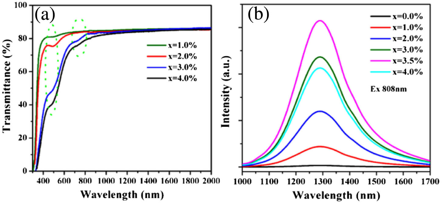
Zaijin Fang1, Síle Nic Chormaic2, Shanyu Wang1, Xin Wang1, Jibo Yu1, Yuxuan Jiang1, Jianrong Qiu3、4, and Pengfei Wang1、5、*
Author Affiliations
  • 1Key Laboratory of In-Fiber Integrated Optics, Ministry Education of China, Harbin Engineering University, Harbin 150001, China
  • 2Light-Matter Interactions Unit, Okinawa Institute of Science and Technology Graduate University, Onna, Okinawa 904-0495, Japan
  • 3State Key Laboratory of Modern Optical Instrumentation, Zhejiang University, Hangzhou 310027, China
  • 4e-mail: qjr@zju.edu.cn
  • 5Photonics Research Centre, Dublin Institute of Technology, Kevin Street, Dublin 8, Ireland
  • show less
    DOI: 10.1364/PRJ.5.000740 Cite this Article Set citation alerts
    Zaijin Fang, Síle Nic Chormaic, Shanyu Wang, Xin Wang, Jibo Yu, Yuxuan Jiang, Jianrong Qiu, Pengfei Wang. Bismuth-doped glass microsphere lasers[J]. Photonics Research, 2017, 5(6): 740 Copy Citation Text show less
    (a) Transmission spectra of xBi2O3-doped germanate glasses (x=1.0, 2.0, 3.0, and 4.0). The different absorption bands around 500 and 800 nm over the transmission spectra are marked by two green dashed elliptical circles. (b) Photoluminescence spectra of xBi2O3-doped germanate glasses under 808 nm excitation (x=0.0, 1.0, 2.0, 3.0, 3.5, and 4.0).
    Fig. 1. (a) Transmission spectra of xBi2O3-doped germanate glasses (x=1.0, 2.0, 3.0, and 4.0). The different absorption bands around 500 and 800 nm over the transmission spectra are marked by two green dashed elliptical circles. (b) Photoluminescence spectra of xBi2O3-doped germanate glasses under 808 nm excitation (x=0.0, 1.0, 2.0, 3.0, 3.5, and 4.0).
    (a) Microscope images of (a) Bi-doped germanate glass fiber and (b) microsphere. (c) XRD patterns of Bi-doped germanate glass, glass fibers and microspheres.
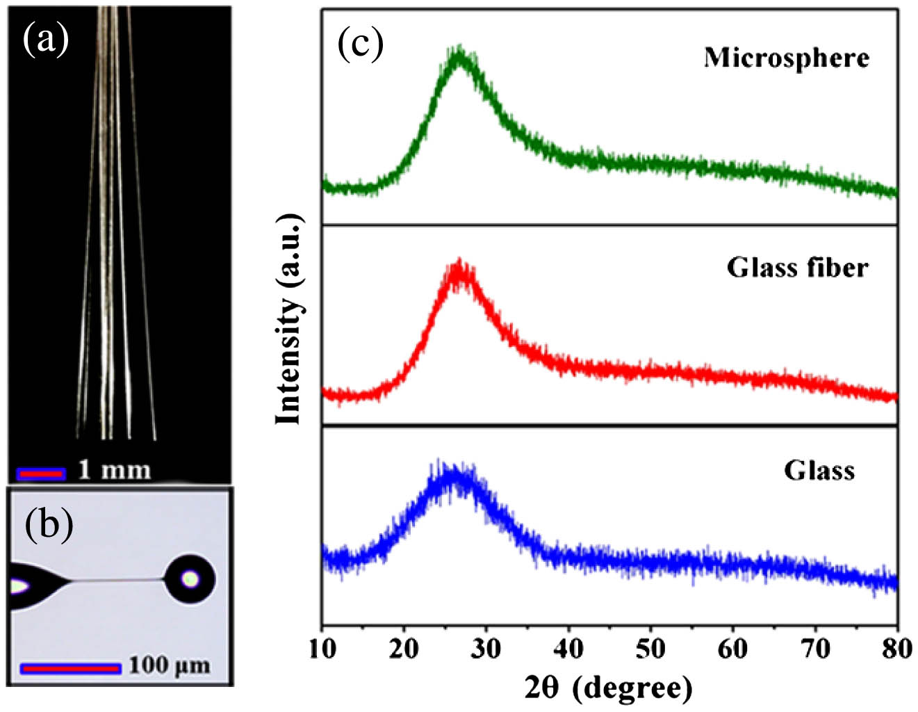
    Fig. 2. (a) Microscope images of (a) Bi-doped germanate glass fiber and (b) microsphere. (c) XRD patterns of Bi-doped germanate glass, glass fibers and microspheres.
    Experimental setup for characterizing a Bi-doped microsphere laser. An 808 nm laser diode is used as the excitation source.
    Fig. 3. Experimental setup for characterizing a Bi-doped microsphere laser. An 808 nm laser diode is used as the excitation source.
    (a) WGMs observed when light is coupled into doped microsphere via the fiber taper coupler. (b) Laser emission (red curve) from the Bi-doped microsphere when the absorbed pump power reaches 215 μW. As a reference, the fluorescence spectrum from the Bi-doped multi-component glass is also shown (blue curve).
    Fig. 4. (a) WGMs observed when light is coupled into doped microsphere via the fiber taper coupler. (b) Laser emission (red curve) from the Bi-doped microsphere when the absorbed pump power reaches 215 μW. As a reference, the fluorescence spectrum from the Bi-doped multi-component glass is also shown (blue curve).
    (a) Microsphere laser output power as a function of estimated absorbed pump power at 1305.8 nm. The straight red line is a linear fit to the experimental data. (b) Oscilloscope trace of the Bi-doped microsphere laser recorded as the pump is scanned in frequency. (c) Schematic of the experimental setup for the linewidth measurement of Bi laser emission.
    Fig. 5. (a) Microsphere laser output power as a function of estimated absorbed pump power at 1305.8 nm. The straight red line is a linear fit to the experimental data. (b) Oscilloscope trace of the Bi-doped microsphere laser recorded as the pump is scanned in frequency. (c) Schematic of the experimental setup for the linewidth measurement of Bi laser emission.
    Zaijin Fang, Síle Nic Chormaic, Shanyu Wang, Xin Wang, Jibo Yu, Yuxuan Jiang, Jianrong Qiu, Pengfei Wang. Bismuth-doped glass microsphere lasers[J]. Photonics Research, 2017, 5(6): 740
    Download Citation