• Laser & Optoelectronics Progress
  • Vol. 60, Issue 12, 1215005 (2023)
Danni Sun, Qibing Zhu*, and Min Huang
Author Affiliations
  • Key Laboratory of Advanced Process Control for Light Industry, Ministry of Education, School of Internet of Things Engineering, Jiangnan University, Wuxi 214122, Jiangsu, China
  • show less
    DOI: 10.3788/LOP221515 Cite this Article Set citation alerts
    Danni Sun, Qibing Zhu, Min Huang. Evaluation of Line-Scan Imaging System's Ability to Detect Internal Defects in Tissue Using Improved Monte Carlo Simulation and Optical Density Algorithm[J]. Laser & Optoelectronics Progress, 2023, 60(12): 1215005 Copy Citation Text show less
    Defective tissue model
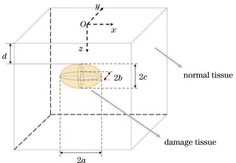
    Fig. 1. Defective tissue model
    Schematic of line-scan virtual imaging system. (a) Line-scan imaging system; (b) line-scan schematic
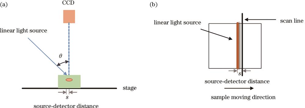
    Fig. 2. Schematic of line-scan virtual imaging system. (a) Line-scan imaging system; (b) line-scan schematic
    Intersection of photon and voxel boundary
    Fig. 3. Intersection of photon and voxel boundary
    Comparison of differences between normal and damaged tissues by OD algorithm along same scan lines. (a) Normal tissue; (b) damage tissue
    Fig. 4. Comparison of differences between normal and damaged tissues by OD algorithm along same scan lines. (a) Normal tissue; (b) damage tissue
    Simulation results of different incident angles of linear light sources
    Fig. 5. Simulation results of different incident angles of linear light sources
    Relationship between diffuse reflectance and source-detector distance
    Fig. 6. Relationship between diffuse reflectance and source-detector distance
    Relationship between source-detector distance and detection depth and photon average path length
    Fig. 7. Relationship between source-detector distance and detection depth and photon average path length
    Optical density differential distribution images. (a) Small defect; (b) large defect
    Fig. 8. Optical density differential distribution images. (a) Small defect; (b) large defect
    CNR change of three defects at different depths
    Fig. 9. CNR change of three defects at different depths
    Optical density difference curves of small defects with different depths
    Fig. 10. Optical density difference curves of small defects with different depths
    Two-dimensional optical density difference of small defects at a depth of 1 mm
    Fig. 11. Two-dimensional optical density difference of small defects at a depth of 1 mm
    ModelTypeParameterValueReference
    Heterogeneous tissueRefractive index n11.35
    Fruit fleshAbsorption coefficient μa1 /cm-10.152313-17
    Scattering coefficient μs1 /cm-14.167
    Anisotropy factor g10.66
    Refractive index n21.365
    Internal defectAbsorption coefficient μa2 /cm-11.63313-17
    Scattering coefficient μs2 /cm-14.564
    Anisotropy factor g20.66
    Table 1. Optical properties of defective tissue model
    ParameterValue
    Number of photons3,000,000
    Resolution of depth (dz);Resolution along x-axis (dx);Resolution along y-axis (dy0.1 mm;0.1 mm;0.1 mm
    Number of grid elements (z,x,y)400;400;400
    Number of tissue types3
    Profile of the incident light beamLinear light source;Gaussian;1-mm width
    Total energy of the incident light beam1 J
    1/e2 radius of the incident light beam0.5 mm
    Table 2. Input parameters for IMC simulation of photon propagation
    Danni Sun, Qibing Zhu, Min Huang. Evaluation of Line-Scan Imaging System's Ability to Detect Internal Defects in Tissue Using Improved Monte Carlo Simulation and Optical Density Algorithm[J]. Laser & Optoelectronics Progress, 2023, 60(12): 1215005
    Download Citation