• Acta Physica Sinica
  • Vol. 69, Issue 10, 102902-1 (2020)
Qian-Li Li1, Ya-Hua Hu2, Yi-Fan Ma1, Zhi-Xiang Sun3, Min Wang1, Xiao-Lin Liu3, Jing-Tai Zhao1, and Zhi-Jun Zhang1、*
Author Affiliations
  • 1School of Materials Science and Engineering, Shanghai University, Shanghai 200444, China
  • 2College of Nanhu, Jiaxing University, Jiaxing 314001, China
  • 3School of Physics Science and Engineering, Tongji University, Shanghai 200092, China
  • show less
    DOI: 10.7498/aps.69.20200282 Cite this Article
    Qian-Li Li, Ya-Hua Hu, Yi-Fan Ma, Zhi-Xiang Sun, Min Wang, Xiao-Lin Liu, Jing-Tai Zhao, Zhi-Jun Zhang. Preparation and properties for X-ray scintillation screen based on ZnO:In nanorod arrays[J]. Acta Physica Sinica, 2020, 69(10): 102902-1 Copy Citation Text show less
    The schematic illustration of the fabrication process of ZnO:In nanorod arrays.
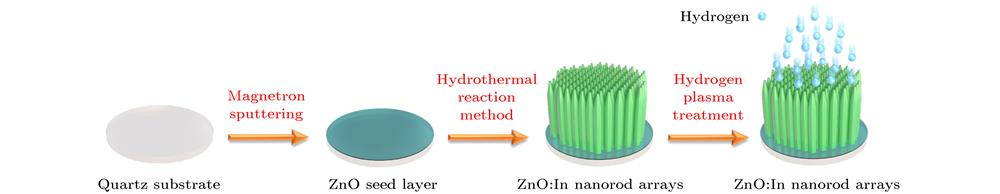
    Fig. 1. The schematic illustration of the fabrication process of ZnO:In nanorod arrays.
    SEM images of ZnO:In nanorod arrays: (a) Cross-sectional; (b) top; (c) surface; (d) oblique views.
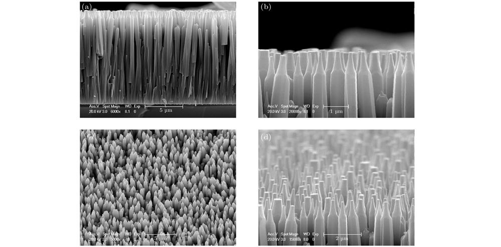
    Fig. 2. SEM images of ZnO:In nanorod arrays: (a) Cross-sectional; (b) top; (c) surface; (d) oblique views.
    XRD patterns of the ZnO:In nanorod arrays before and after hydrogen plasma treatment.
    Fig. 3. XRD patterns of the ZnO:In nanorod arrays before and after hydrogen plasma treatment.
    XEL spectra of the ZnO:In nanorod arrays before and after hydrogen plasma treatment.
    Fig. 4. XEL spectra of the ZnO:In nanorod arrays before and after hydrogen plasma treatment.
    The fluorescence decay curves of (a) ultravioletemission (λex = 325 nm, λem = 395 nm) and (b) visible emission (λex = 325 nm, λem = 530 nm) for the ZnO:In nanorod arrays.
    Fig. 5. The fluorescence decay curves of (a) ultravioletemission (λex = 325 nm, λem = 395 nm) and (b) visible emission (λex = 325 nm, λem = 530 nm) for the ZnO:In nanorod arrays.
    Schematic diagram of the synchrotron radiation X-ray imaging measurement setup at BL13 W1, SSRF.
    Fig. 6. Schematic diagram of the synchrotron radiation X-ray imaging measurement setup at BL13 W1, SSRF.
    (a) Physical, Schematic diagram of internal structure and Micron-resolved pattern of JIMA RT-02 micro-resolution plates; the X-ray images of (b) 3 μm and (c) 1.5 μm basedonZnO:In nanorod arrays as the scintillation screen.
    Fig. 7. (a) Physical, Schematic diagram of internal structure and Micron-resolved pattern of JIMA RT-02 micro-resolution plates; the X-ray images of (b) 3 μm and (c) 1.5 μm basedonZnO:In nanorod arrays as the scintillation screen.
    (a) MTF and (b) DQE curves of the X-ray imaging system with ZnO:In nanorod arrays.
    Fig. 8. (a) MTF and (b) DQE curves of the X-ray imaging system with ZnO:In nanorod arrays.
    Qian-Li Li, Ya-Hua Hu, Yi-Fan Ma, Zhi-Xiang Sun, Min Wang, Xiao-Lin Liu, Jing-Tai Zhao, Zhi-Jun Zhang. Preparation and properties for X-ray scintillation screen based on ZnO:In nanorod arrays[J]. Acta Physica Sinica, 2020, 69(10): 102902-1
    Download Citation