
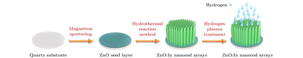
Qian-Li Li, Ya-Hua Hu, Yi-Fan Ma, Zhi-Xiang Sun, Min Wang, Xiao-Lin Liu, Jing-Tai Zhao, Zhi-Jun Zhang. Preparation and properties for X-ray scintillation screen based on ZnO:In nanorod arrays [J]. Acta Physica Sinica, 2020, 69(10): 102902-1
- Acta Physica Sinica
- Vol. 69, Issue 10, 102902-1 (2020)
Abstract


Set citation alerts for the article
Please enter your email address

© Copyright 2018-2021 | Chinese Laser Press. All Rights Reserved 沪ICP备15018463号-20