• Acta Optica Sinica
  • Vol. 43, Issue 5, 0511002 (2023)
Luhan Wang1、2, Yan Gong2、3、*, Yanwei Zhang2, Song Lang2、4, and Hanqing Zheng2
Author Affiliations
  • 1Life Sciences and Medicine, School of Biomedical Engineering (Suzhou), University of Science and Technology of China, Suzhou 215163, Jiangsu, China
  • 2Medical Optical Research Laboratory, Suzhou Institute of Biomedical Engineering Technology, Chinese Academy of Sciences, Suzhou 215163, Jiangsu, China
  • 3Jinan Guoke Medical Technology Development Co., Ltd., Jinan 250102, Shandong, China
  • 4Shenyang Guoke Medical Technology Development Co., Ltd., Shenyang 110167, Liaoning, China
  • show less
    DOI: 10.3788/AOS221703 Cite this Article Set citation alerts
    Luhan Wang, Yan Gong, Yanwei Zhang, Song Lang, Hanqing Zheng. Human Eye Autofocus and Pupil Center Auto-Alignment System[J]. Acta Optica Sinica, 2023, 43(5): 0511002 Copy Citation Text show less
    Structure of human eye auto-focus and pupil center auto-alignment device
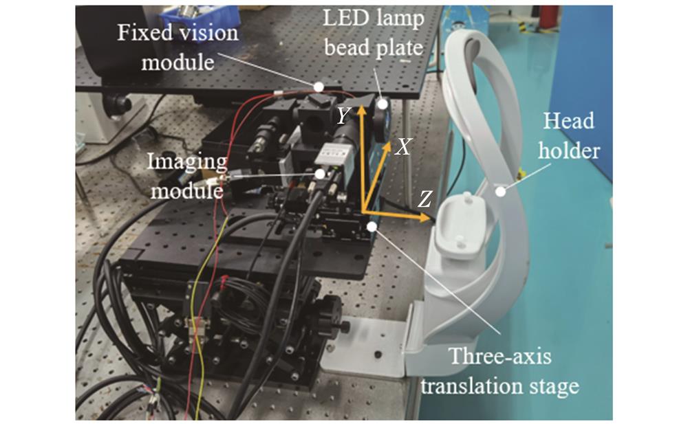
    Fig. 1. Structure of human eye auto-focus and pupil center auto-alignment device
    Schematic of optical path of imaging module
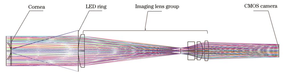
    Fig. 2. Schematic of optical path of imaging module
    Flowchart of pupil center positioning and alignment algorithm
    Fig. 3. Flowchart of pupil center positioning and alignment algorithm
    Collected image of human eye and ROI image. (a) Original image of human eye; (b) ROI image of pupil
    Fig. 4. Collected image of human eye and ROI image. (a) Original image of human eye; (b) ROI image of pupil
    Grayscale histogram distributions before and after extraction of ROI. (a) Grayscale histogram distribution of original image; (b) grayscale histogram distribution of ROI
    Fig. 5. Grayscale histogram distributions before and after extraction of ROI. (a) Grayscale histogram distribution of original image; (b) grayscale histogram distribution of ROI
    ROI image after threshold segmentation. (a) Image after T1 threshold segmentation; (b) image after T2 threshold segmentation
    Fig. 6. ROI image after threshold segmentation. (a) Image after T1 threshold segmentation; (b) image after T2 threshold segmentation
    Schematic diagrams of pupil center and optical axis center. (a) Pupil identification and center point; (b) optical axis ellipse fitting and center point
    Fig. 7. Schematic diagrams of pupil center and optical axis center. (a) Pupil identification and center point; (b) optical axis ellipse fitting and center point
    Schematic diagram of relative position of pupil center and optical axis
    Fig. 8. Schematic diagram of relative position of pupil center and optical axis
    Schematic diagrams of defocus sequence-1 of human eyes. (a) 5th image of sequence; (b) 9th image of sequence; (c) 13th image of sequence; (d) 17th image of sequence; (e) 21st image of sequence
    Fig. 9. Schematic diagrams of defocus sequence-1 of human eyes. (a) 5th image of sequence; (b) 9th image of sequence; (c) 13th image of sequence; (d) 17th image of sequence; (e) 21st image of sequence
    Normalized sharpness evaluation curves of human eye defocused image sequences. (a) Sequence 1; (b) sequence 2;(c) sequence 3; (d) sequence 4
    Fig. 10. Normalized sharpness evaluation curves of human eye defocused image sequences. (a) Sequence 1; (b) sequence 2;(c) sequence 3; (d) sequence 4
    Partial images of sequence 4 in human eye defocused image. (a) 8th image of sequence; (b) 9th image of sequence
    Fig. 11. Partial images of sequence 4 in human eye defocused image. (a) 8th image of sequence; (b) 9th image of sequence
    Error between pupil center coordinates obtained by three algorithms and manually calibrated pupil center coordinates
    Fig. 12. Error between pupil center coordinates obtained by three algorithms and manually calibrated pupil center coordinates
    Processing results of algorithm in this paper under complex conditions. (a) Dark light condition; (b) bright light condition; (c)(d) partial occlusion of pupil
    Fig. 13. Processing results of algorithm in this paper under complex conditions. (a) Dark light condition; (b) bright light condition; (c)(d) partial occlusion of pupil
    Evaluation functionTPF /msRFsenUnimodalityAccuracy
    FMTG13.093.5310.329
    Brenner14.12.8000.188
    Tenengrad42.81.5610.114
    SMD16.11.1380.058××
    Laplace13.81.1100.026××
    Vollaths15.71.2040.050××
    Variance15.51.2780.053××
    Table 1. Average performance indexes of evaluation curves of out-of-focus sequences 1-4 of human eye images
    MethodAverage time /msAccuracy /%Average error /pixel
    LPC10.297.141.4
    Improved Hough584.753.333.3
    Hough5100.414.298.4
    Table 2. Average computation time, accuracy, and average error of three algorithms under experimental images
    Luhan Wang, Yan Gong, Yanwei Zhang, Song Lang, Hanqing Zheng. Human Eye Autofocus and Pupil Center Auto-Alignment System[J]. Acta Optica Sinica, 2023, 43(5): 0511002
    Download Citation