
Keyu Chen, Limin Xu, Jie Gan, Shifeng Wen, Yan Zhou. Effects of Laser Power on Microstructure and Mechanical Properties of Selective Laser Melted AlSi10Mg[J]. Laser & Optoelectronics Progress, 2021, 58(13): 1314001
- Laser & Optoelectronics Progress
- Vol. 58, Issue 13, 1314001 (2021)
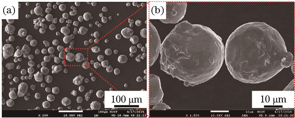
Fig. 1. Morphologies of AlSi10Mg powder
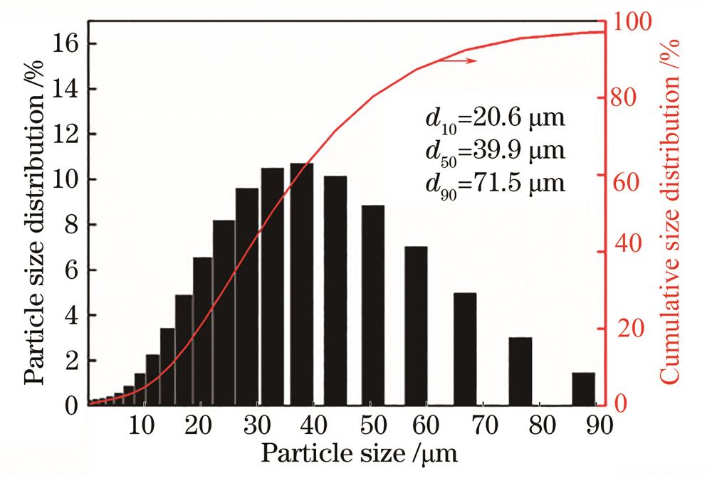
Fig. 2. Particle distribution of AlSi10Mg powder
Fig. 3. Scanning strategy and size of tensile samples. (a) Scanning strategy; (b) size of tensile sample
Fig. 4. XRD spectra of AlSi10Mg samples formed at different laser powers. (a) 20°~90°;(b) 36°~48°
Fig. 5. Melting tracks on the upper surface of AlSi10Mg samples formed at different laser powers. (a) 250 W; (b) 300 W; (c) 350 W; (d) 400 W
Fig. 6. Optical micrographs of the surface of AlSi10Mg samples formed at different laser powers. (a) 250 W; (b) 300 W; (c) 350 W; (d) 400 W
Fig. 7. Relative density of AlSi10Mg samples formed at different laser powers
Fig. 8. Microstructures of AlSi10Mg sample formed by SLM. (a) Microstructure at low magnification; (b) fine grained region; (c) heat affected zone; (d) coarse grained region
Fig. 9. Energy spectrum analysis. (a)‒(c) Energy spectra of white network phase and gray-black matrix; (d) elements contents obtained by point-scanning
Fig. 10. Fine grained region at different laser powers. (a) 250 W; (b) 300 W; (c) 350 W; (d) 400 W
Fig. 11. Load-indentation depth curves of AlSi10Mg samples formed at different laser powers
Fig. 12. Variation of micro-hardness of AlSi10Mg samples with laser power
Fig. 13. Tensile stress-strain curves of AlSi10Mg samples formed at different laser powers
Fig. 14. Tensile strength and elongation of AlSi10Mg samples formed at different laser powers
Fig. 15. Tensile fracture morphologies of AlSi10Mg sample

Set citation alerts for the article
Please enter your email address
© Copyright 2018-2021 | Chinese Laser Press. All Rights Reserved 沪ICP备15018463号-20