
Dantong Zhu, Honghai Shen, Mingyu Yang, Cheng Chen, Tongling Nan. Analysis and Correction of Coded Pixel Distortion in Coded Aperture Imaging Spectrometer[J]. Laser & Optoelectronics Progress, 2018, 55(6): 061201
- Laser & Optoelectronics Progress
- Vol. 55, Issue 6, 061201 (2018)
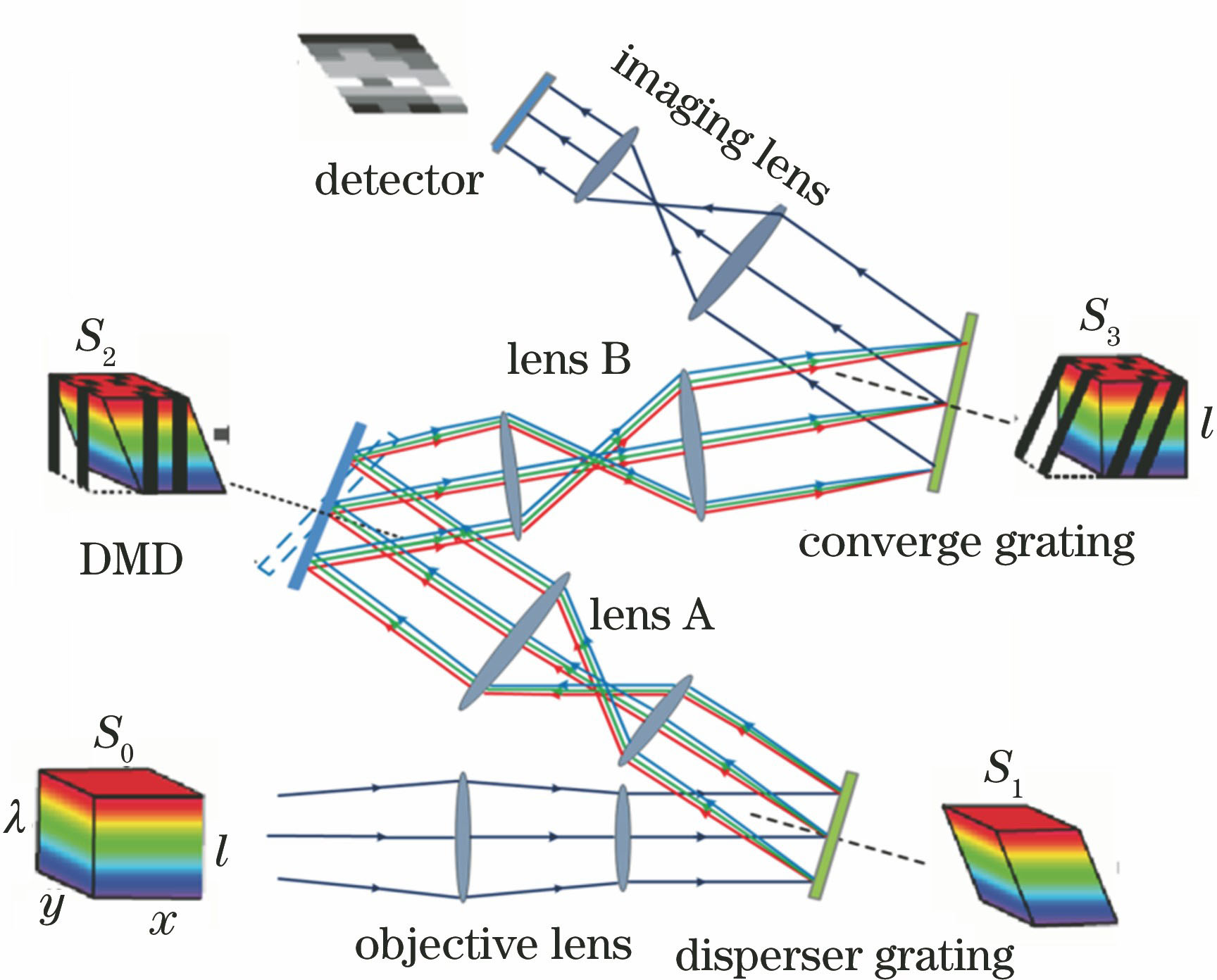
Fig. 1. Principle of SDCAIS based on DMD
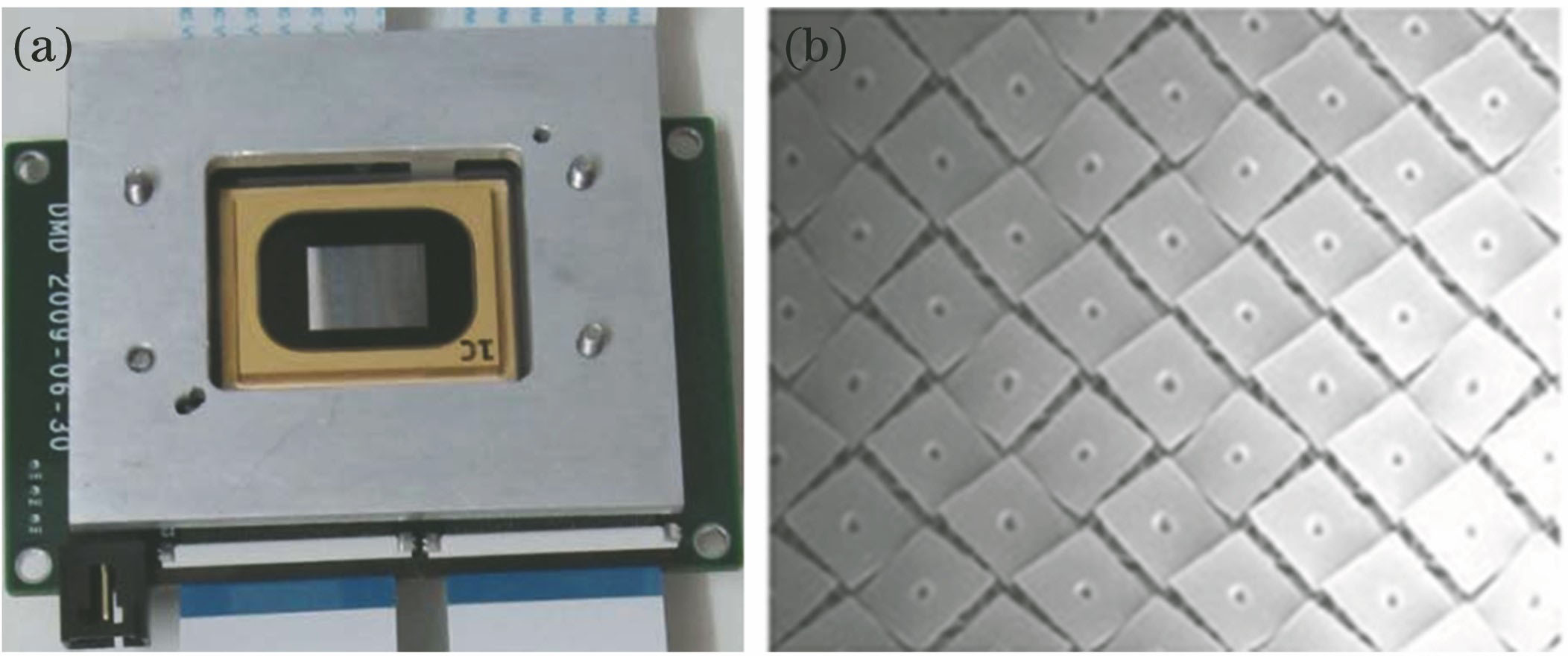
Fig. 2. (a) Appearance and (b) reflective array of DMD
Fig. 3. Correspondence of DMD and the detector in space
Fig. 4. Correspondence of DMD and the detector. (a) In ideal condition; (b) in fact condition
Fig. 5. Optical path of the experiment
Fig. 6. Image which is out of focus
Fig. 7. Hollow square calibration pattern with the fringe width of 8 pixel loaded on DMD
Fig. 8. Image after matching for detector and DMD in the direction of θ
Fig. 9. Location of the center of DMD on detector
Fig. 10. Comparison diagram of the effect. (a) Effect of mean value; (b) detail of mean value; (c) effect of mid-value; (d) detail of mid-value
Fig. 11. Result after binarizational processing
Fig. 12. Results after processing with each edge extraction operator. (a) Prewitt operator: threshold is 0.04; (b) Log operator: threshold is 2; (c) Log operator: threshold is 3; (d) Canny operator: threshold is 0.2
Fig. 13. Schematic diagram of tilt angle
Fig. 14. Comparison of before and after correction. (a) Before correction; (b) after correction
Fig. 15. Pattern collected by detector in theory
Fig. 16. Comparison of the hollow square pattern with the fringe width of 4 pixel (a) before and (b) after correction
Fig. 17. Grid stripes with the side length of 8 pixel (a) before and (b) after correction; grid stripes with the side length of 16 pixel (c) before and (d) after correction
Fig. 18. Colorful experiment objective
Fig. 19. Comparison of colorful dull image with 15 orders coding before and after correction. (a) Before correction; (b) after correction
Fig. 20. Image after 2D mid-value filtering process

Set citation alerts for the article
Please enter your email address
© Copyright 2018-2021 | Chinese Laser Press. All Rights Reserved 沪ICP备15018463号-20