
Chenying Zhang, Wei Zhou, Da Geng, Cheng Bai, Weida Li, Songyue Chen, Tao Luo, Lifeng Qin, Yu Xie. Laser direct writing and characterizations of flexible piezoresistive sensors with microstructures[J]. Opto-Electronic Advances, 2021, 4(4): 200061-1
- Opto-Electronic Advances
- Vol. 4, Issue 4, 200061-1 (2021)
Abstract
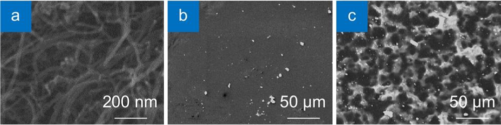
Figure S1 shows the morphologies of MWCNTs, PDMS and MPC. As shown in Fig. S1(a), MWCNTs show a high length-diameter ratio and get tangled up with each other. It is attributed to the Van der Waals force between MWCNTs. Consequently, MPC show much rougher surface than pure PDMS due to the internal agglomeration of MWCNTs, as shown in Fig. S1(b) and Fig. S1(c).
The mechanical evaluation system is shown in Fig. S2.
The strain along stretching direction (εz) is calculated as Eq. S1:
where
On the cross section of the testing sample, the width w and thickness t direction (εx) are calculated as Eq. S2:
where w0 and
The tensile stress (σz) is calculated as Eq. S3:
where
The relationships of εz and σz can be described by Young's modulus (E), as shown in Eq. S4:
Consequently, E of testing samples can be evaluated by slopes of σz-εz curves.
Figure S3 shows the variation of laser power and pulse with repetition frequency at Q = 1. Laser pulse is increased with the increase of repetition frequency. When repetition frequency is below 80 kHz, laser power increases with the increasing repetition frequency. When repetition frequency exceeds 90 kHz, laser power decreases with the increasing repetition frequency.
Figure S4 shows surface morphologies of the microstructures fabricated with different laser parameters. When laser repetition frequency varied from 35 kHz to 45 kHz, laser power varied in an approximate range of 2~5 W, which brought about significant impacts in morphology of microstructures. When laser scanning speed varied from 100 mm·s-1 to 200 mm·s-1, obvious difference in morphology of the microstructures could be found, which can be attributed to the significant variation in the laser energy density. When laser repetition frequency was below 40 kHz, laser path can be found on the substrates and the microstructures had relative small size. When laser repetition frequency was set to 45 kHz, the laser path disappeared from substrates because a higher energy density induced significant ablation and quantities of particles can be found around the microstructures, which indicated destruction to the microstructures.
Figure S5 shows sensitivity of flexible piezoresistive sensors fabricated by different laser parameters. Laser parameters significantly affected the sensitivity and the performance of flexible piezoresistive sensors through modulating the surface morphologies of microstructures.
References
[1] YN Ma, NS Liu, LY Li, XK Hu, ZG Zou, et al. A highly flexible and sensitive piezoresistive sensor based on MXene with greatly changed interlayer distances. Nat Commun, 8, 1207(2017).
[2] L Zhang, HQ Li, XJ Lai, TY Gao, J Yang, et al. Thiolated Graphene@Polyester Fabric-Based Multilayer Piezoresistive Pressure Sensors for Detecting Human Motion. ACS Appl Mater Interfaces, 10, 41784-41792(2018).
[3] GY Bae, SW Pak, D Kim, G Lee, D Kim, et al. Linearly and Highly Pressure-Sensitive Electronic Skin Based on a Bioinspired Hierarchical Structural Array. Adv Mater, 28, 5300(2016).
[4] L Liu, Y Huang, FY Li, Y Ma, WB Li, et al. Spider-web inspired multi-resolution graphene tactile sensor. Chem Commun, 54, 4810-4813(2018).
[5] J Wang, M Tenjimbayashi, Y Tokura, JY Park, K Kawase, et al. Bionic Fish-Scale Surface Structures Fabricated via Air/Water Interface for Flexible and Ultrasensitive Pressure Sensors. ACS Appl Mater Interfaces, 10, 30689-30697(2018).
[6] Z Wang, X Guan, H Huang, H Wang, W Lin, et al. Full 3D Printing of Stretchable Piezoresistive Sensor with Hierarchical Porosity and Multimodulus Architecture. Adv Funct Mater, 29, 1807569(2019).
[7] Y Pang, KN Zhang, Z Yang, S Jiang, ZY Ju, et al. Epidermis Microstructure Inspired Graphene Pressure Sensor with Random Distributed Spinosum for High Sensitivity and Large Linearity. ACS Nano, 12, 2346-2354(2018).
[8] TH Chang, Y Tian, CS Li, XY Gu, KR Li, et al. Stretchable Graphene Pressure Sensors with Shar-Pei-like Hierarchical Wrinkles for Collision-Aware Surgical Robotics. ACS Appl Mater Interfaces, 11, 10226-10236(2019).

Set citation alerts for the article
Please enter your email address
© Copyright 2018-2021 | Chinese Laser Press. All Rights Reserved 沪ICP备15018463号-20