• Laser & Optoelectronics Progress
  • Vol. 61, Issue 2, 0211025 (2024)
Dejun Liu1、2、†, Ziyi Huang1、2、†, Zhuorong Li1、2, Yalong Tai1、2, Xiaobin Wang3, Li Zhang4, Changrui Liao1、2、*, and Yiping Wang1、2
Author Affiliations
  • 1Key Laboratory of Optoelectronic Devices and Systems of Ministry of Education/Guangdong Province, Shenzhen Key Laboratory of Ultrafast Laser Micro/Nano Manufacturing, College of Physics and Optoelectronic Engineering, Shenzhen University, Shenzhen 518060, Guangdong , China
  • 2State Key Laboratory of Radio Frequency Heterogeneous Integration, Guangdong and HongKong Joint Research Centre for Optical Fibre Sensors, Shenzhen Key Laboratory of Photonic Devices and Sensing Systems for Internet of Things, Shenzhen University, Shenzhen 518060, Guangdong , China
  • 3Department of Urology, Southern University of Science and Technology Hospital, Shenzhen 518055, Guangdong , China
  • 4Department of Mechanical and Automation Engineering, The Chinese University of Hong Kong, Shatin NT, 999077, Hong Kong SAR , China
  • show less
    DOI: 10.3788/LOP232208 Cite this Article Set citation alerts
    Dejun Liu, Ziyi Huang, Zhuorong Li, Yalong Tai, Xiaobin Wang, Li Zhang, Changrui Liao, Yiping Wang. Recent Advances in Micro-Endoscopies Based on Optical Coherence Tomography (Invited)[J]. Laser & Optoelectronics Progress, 2024, 61(2): 0211025 Copy Citation Text show less
    Comparison of resolution among different imaging technologies[3]
    Fig. 1. Comparison of resolution among different imaging technologies[3]
    Configurations of fiber OCT systems. (a) TD-OCT; (b) SD-OCT; (c) SS-OCT
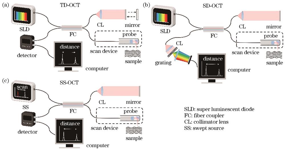
    Fig. 2. Configurations of fiber OCT systems. (a) TD-OCT; (b) SD-OCT; (c) SS-OCT
    Fiber OCT imaging catheters. (a) Forward-viewing catheter; (b) side-viewing catheter; (c) proximal scanning catheter; (d) distal scanning catheter
    Fig. 3. Fiber OCT imaging catheters. (a) Forward-viewing catheter; (b) side-viewing catheter; (c) proximal scanning catheter; (d) distal scanning catheter
    Fiber-prism-based fiber OCT endoscopes. (a) Catheter type[21]; (b) needle type[24]; (c) balloon type[29]; (d) capsule type[30]
    Fig. 4. Fiber-prism-based fiber OCT endoscopes. (a) Catheter type[21]; (b) needle type[24]; (c) balloon type[29]; (d) capsule type[30]
    Design of fiber OCT endoscope with high resolution and long depth of focus. (a) High resolution and long depth of focus achieved with diffractive lens[32]; (b) long depth of focus imaging achieved by self-imaging wavefront division[34]
    Fig. 5. Design of fiber OCT endoscope with high resolution and long depth of focus. (a) High resolution and long depth of focus achieved with diffractive lens[32]; (b) long depth of focus imaging achieved by self-imaging wavefront division[34]
    All-fiber OCT endoscopes. (a) Forward-viewing fiber OCT endoscope based on GRIN fiber[38]; (b) side-viewing fiber OCT endoscope based on angle polished ball lens[40]
    Fig. 6. All-fiber OCT endoscopes. (a) Forward-viewing fiber OCT endoscope based on GRIN fiber[38]; (b) side-viewing fiber OCT endoscope based on angle polished ball lens[40]
    Design of all-fiber OCT endoscope with long depth of focus. (a) Endoscope with phase mask[43]; (b) axicon lens[44]; (c) no-focus collimated light beam[45]
    Fig. 7. Design of all-fiber OCT endoscope with long depth of focus. (a) Endoscope with phase mask[43]; (b) axicon lens[44]; (c) no-focus collimated light beam[45]
    Composite fiber OCT endoscopes. (a) BPSF endoscope[46]; (b) metalens endoscope[48]
    Fig. 8. Composite fiber OCT endoscopes. (a) BPSF endoscope[46]; (b) metalens endoscope[48]
    Composite fiber OCT endoscopes based on two-photon polymerization 3D printing. (a) Separate probe[56]; (b) monolithic probe[57]
    Fig. 9. Composite fiber OCT endoscopes based on two-photon polymerization 3D printing. (a) Separate probe[56]; (b) monolithic probe[57]
    Endoscopic imaging of human lung resections using OCT probe based on metalens[48], the fine features of lung tissue are clearly visible in the magnified OCT images, including moderately scattering epithelium (epi), highly scattering basement membrane (bm), cartilage (car), alveoli (alv), and the small irregular glands (g). (a) Normal airway; (b) distal bronchiole; (c) abnormal airway
    Fig. 10. Endoscopic imaging of human lung resections using OCT probe based on metalens[48], the fine features of lung tissue are clearly visible in the magnified OCT images, including moderately scattering epithelium (epi), highly scattering basement membrane (bm), cartilage (car), alveoli (alv), and the small irregular glands (g). (a) Normal airway; (b) distal bronchiole; (c) abnormal airway
    OCT images of esophagus of different animals and their histological images of the specimen. (a) Swine esophagus[34]. Images obtained with the conventional Gauss beam (1) are significantly blurred when it is out of focus, while the cells are visualized with high contrast and resolution in images obtained with the CAFM beam (2) due to longer depth of focus; (b) rat esophagus[40]. The cut-away view of a reconstructed 3D image (1) and cross-sectional image (2) obtained by OCT scanning, where the keratinized stratified squamous epithelium (EP), lamina propria (LP), muscularis mucosae (MM), muscularis propria (MP), and submucosa (SM) are observed in enlarged view
    Fig. 11. OCT images of esophagus of different animals and their histological images of the specimen. (a) Swine esophagus[34]. Images obtained with the conventional Gauss beam (1) are significantly blurred when it is out of focus, while the cells are visualized with high contrast and resolution in images obtained with the CAFM beam (2) due to longer depth of focus; (b) rat esophagus[40]. The cut-away view of a reconstructed 3D image (1) and cross-sectional image (2) obtained by OCT scanning, where the keratinized stratified squamous epithelium (EP), lamina propria (LP), muscularis mucosae (MM), muscularis propria (MP), and submucosa (SM) are observed in enlarged view
    White light endoscopic images and OCT images of esophagus of the colon model[75]. (a) Healthy tissue with layered structure of mucosa (M), submucosa (S), and muscular layer (ML); (b) non neoplastic mucosal growth phantom. Yellow arrows represent a benign lesion with visible thickening of the mucosa; (c) pedunculated polyp phantom; (d) flat cancerous tissue (CT) and healthy tissue (HT) sessile
    Fig. 12. White light endoscopic images and OCT images of esophagus of the colon model[75]. (a) Healthy tissue with layered structure of mucosa (M), submucosa (S), and muscular layer (ML); (b) non neoplastic mucosal growth phantom. Yellow arrows represent a benign lesion with visible thickening of the mucosa; (c) pedunculated polyp phantom; (d) flat cancerous tissue (CT) and healthy tissue (HT) sessile
    Endoscopic OCT images of porcine ureter[81], urothelium (U), lamina propria (LP), smooth muscle (SM), and adipose tissue (AD) are observed with high resolution. (a) Cross-sectional image; (b) histologic image; (c) 3D renderings of ureter segment consisting of multiple cross-sectional images; (d) longitudinal lumen view
    Fig. 13. Endoscopic OCT images of porcine ureter[81], urothelium (U), lamina propria (LP), smooth muscle (SM), and adipose tissue (AD) are observed with high resolution. (a) Cross-sectional image; (b) histologic image; (c) 3D renderings of ureter segment consisting of multiple cross-sectional images; (d) longitudinal lumen view
    OCT scanning images of different arteries. (a) Human cadaver coronary artery[35]. The smooth muscle cells (red arrow) and macrophages undergoing diapedesis (green arrow) are observed in image (1), image (2) shows probable thrombus (blue arrow); (b) mouse aorta[57]. The adventitial and perivascular adipose tissues (AT) in image (1) and cholesterol crystals (yellow arrow) in image (2) are observed
    Fig. 14. OCT scanning images of different arteries. (a) Human cadaver coronary artery[35]. The smooth muscle cells (red arrow) and macrophages undergoing diapedesis (green arrow) are observed in image (1), image (2) shows probable thrombus (blue arrow); (b) mouse aorta[57]. The adventitial and perivascular adipose tissues (AT) in image (1) and cholesterol crystals (yellow arrow) in image (2) are observed
    3D reconstructed OCT images of arteries with implanted stents. (a) Rabbit artery[35]. The purple and red arrows indicate the implanted stents; (b) porcine artery[45]. The blood vessel wall (H), guidewire (G), and stents (S) are observed
    Fig. 15. 3D reconstructed OCT images of arteries with implanted stents. (a) Rabbit artery[35]. The purple and red arrows indicate the implanted stents; (b) porcine artery[45]. The blood vessel wall (H), guidewire (G), and stents (S) are observed
    Micro-OCT system with long depth of focus and fiber probe based on few mode interferometry[35]
    Fig. 16. Micro-OCT system with long depth of focus and fiber probe based on few mode interferometry[35]
    Fiber OCT probes for multimodal imaging. (a) Ultrasound/OCT[98]; (b) photoacoustic/ultrasound/OCT[99]
    Fig. 17. Fiber OCT probes for multimodal imaging. (a) Ultrasound/OCT[98]; (b) photoacoustic/ultrasound/OCT[99]
    Fiber OCT probes for multimodal imaging. (a) Fluorescence/OCT[100]; (b) polarization-sensitive OCT[101]
    Fig. 18. Fiber OCT probes for multimodal imaging. (a) Fluorescence/OCT[100]; (b) polarization-sensitive OCT[101]
    Dejun Liu, Ziyi Huang, Zhuorong Li, Yalong Tai, Xiaobin Wang, Li Zhang, Changrui Liao, Yiping Wang. Recent Advances in Micro-Endoscopies Based on Optical Coherence Tomography (Invited)[J]. Laser & Optoelectronics Progress, 2024, 61(2): 0211025
    Download Citation