• Journal of the European Optical Society-Rapid Publications
  • Vol. 18, Issue 2, 2022011 (2022)
Myeongseop Kim1、#, Hee Ryung Lee1、#, Razvigor Ossikovski1, Aude Malfait-Jobart2, Dominique Lamarque3, and Tatiana Novikova1、4、*
Author Affiliations
  • 1LPICM, CNRS, Ecole Polytechnique, IP Paris, LPICM, Palaiseau 91128, France
  • 2Université Paris-Saclay, Université de Versailles St. Quentin-en-Yvelines, INSERM (UMR 1173), Montigny-Le-Bretonneux, France
  • 3Université de Versailles St-Quentin en Yvelines, Hôpital Ambroise Paré, Service d’Hépato-Gastroentérologie, Boulogne Billancourt, France
  • 4Florida International University, Department of Biomedical Engineering, Miami, FL, USA
  • show less
    DOI: 10.1051/jeos/2022011 Cite this Article
    Myeongseop Kim, Hee Ryung Lee, Razvigor Ossikovski, Aude Malfait-Jobart, Dominique Lamarque, Tatiana Novikova. Optical diagnosis of gastric tissue biopsies with Mueller microscopy and statistical analysis[J]. Journal of the European Optical Society-Rapid Publications, 2022, 18(2): 2022011 Copy Citation Text show less
    White light microscopy images of H&E stained thin sections of three different human gastric endoscopy biopsies: (a) healthy control (c) chronic gastritis; (e) gastric cancer. The insets (b), (d) and (f) show the enlarged black box zones of the images (a), (b) and (e), respectively. The scale bar is 200 μm in (a), (c) and (e) and 100 μm in (b), (d) and (f).
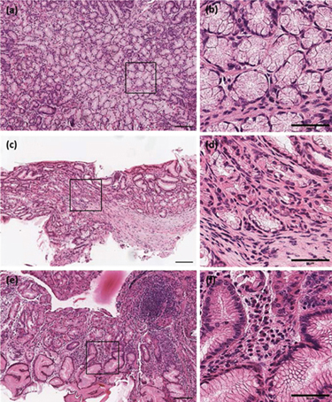
    Fig. 1. White light microscopy images of H&E stained thin sections of three different human gastric endoscopy biopsies: (a) healthy control (c) chronic gastritis; (e) gastric cancer. The insets (b), (d) and (f) show the enlarged black box zones of the images (a), (b) and (e), respectively. The scale bar is 200 μm in (a), (c) and (e) and 100 μm in (b), (d) and (f).
    Mueller microscopy images of unstained thin sections of three different human gastric endoscopy biopsies. Normalized transmitted intensity – (a) healthy control, (c) chronic gastritis; (e) gastric cancer. Map of scalar retardance – (b) healthy control, (d) chronic gastritis; (f) gastric cancer. Field of view is about 250 μm in diameter.
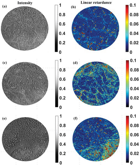
    Fig. 2. Mueller microscopy images of unstained thin sections of three different human gastric endoscopy biopsies. Normalized transmitted intensity – (a) healthy control, (c) chronic gastritis; (e) gastric cancer. Map of scalar retardance – (b) healthy control, (d) chronic gastritis; (f) gastric cancer. Field of view is about 250 μm in diameter.
    Selection of the region of interest (red box – 400 × 300 pixels, left panel) and corresponding training dataset (size of 4 × 400 × 300) – the images of transmitted intensity, linear retardance, linear dichroism, and circular depolarization of healthy control gastric tissue.
    Fig. 3. Selection of the region of interest (red box – 400 × 300 pixels, left panel) and corresponding training dataset (size of 4 × 400 × 300) – the images of transmitted intensity, linear retardance, linear dichroism, and circular depolarization of healthy control gastric tissue.
    The main steps of building the regression model using training set of the intensity and polarimetric images of healthy control gastric tissue section.
    Fig. 4. The main steps of building the regression model using training set of the intensity and polarimetric images of healthy control gastric tissue section.
    Predicted images (400 × 300 pixels) of gastric tissue sections: (a) healthy, (b) chronic gastritis, (c) gastric cancer), (d–f) histograms of the corresponding predicted images; (g–i) insets with the enlarged view of the second Gaussian peak.
    Fig. 5. Predicted images (400 × 300 pixels) of gastric tissue sections: (a) healthy, (b) chronic gastritis, (c) gastric cancer), (d–f) histograms of the corresponding predicted images; (g–i) insets with the enlarged view of the second Gaussian peak.
    Myeongseop Kim, Hee Ryung Lee, Razvigor Ossikovski, Aude Malfait-Jobart, Dominique Lamarque, Tatiana Novikova. Optical diagnosis of gastric tissue biopsies with Mueller microscopy and statistical analysis[J]. Journal of the European Optical Society-Rapid Publications, 2022, 18(2): 2022011
    Download Citation