• Laser & Optoelectronics Progress
  • Vol. 57, Issue 24, 241014 (2020)
Xiaofang Zhu, Liang Jing, and Dangguo Shao*
Author Affiliations
  • Faculty of Information Engineering and Automation, Kunming University of Science and Technology, Kunming, Yunnan 650500, China
  • show less
    DOI: 10.3788/LOP57.241014 Cite this Article Set citation alerts
    Xiaofang Zhu, Liang Jing, Dangguo Shao. Ultrasonic Image Denoising Using Adaptive Bilateral Filtering Based on Back Propagation Neural Network[J]. Laser & Optoelectronics Progress, 2020, 57(24): 241014 Copy Citation Text show less
    Structural diagram of BP neural network
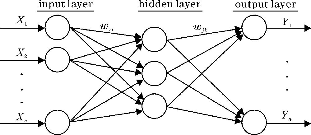
    Fig. 1. Structural diagram of BP neural network
    BP neural network based adaptive bilateral filtering model
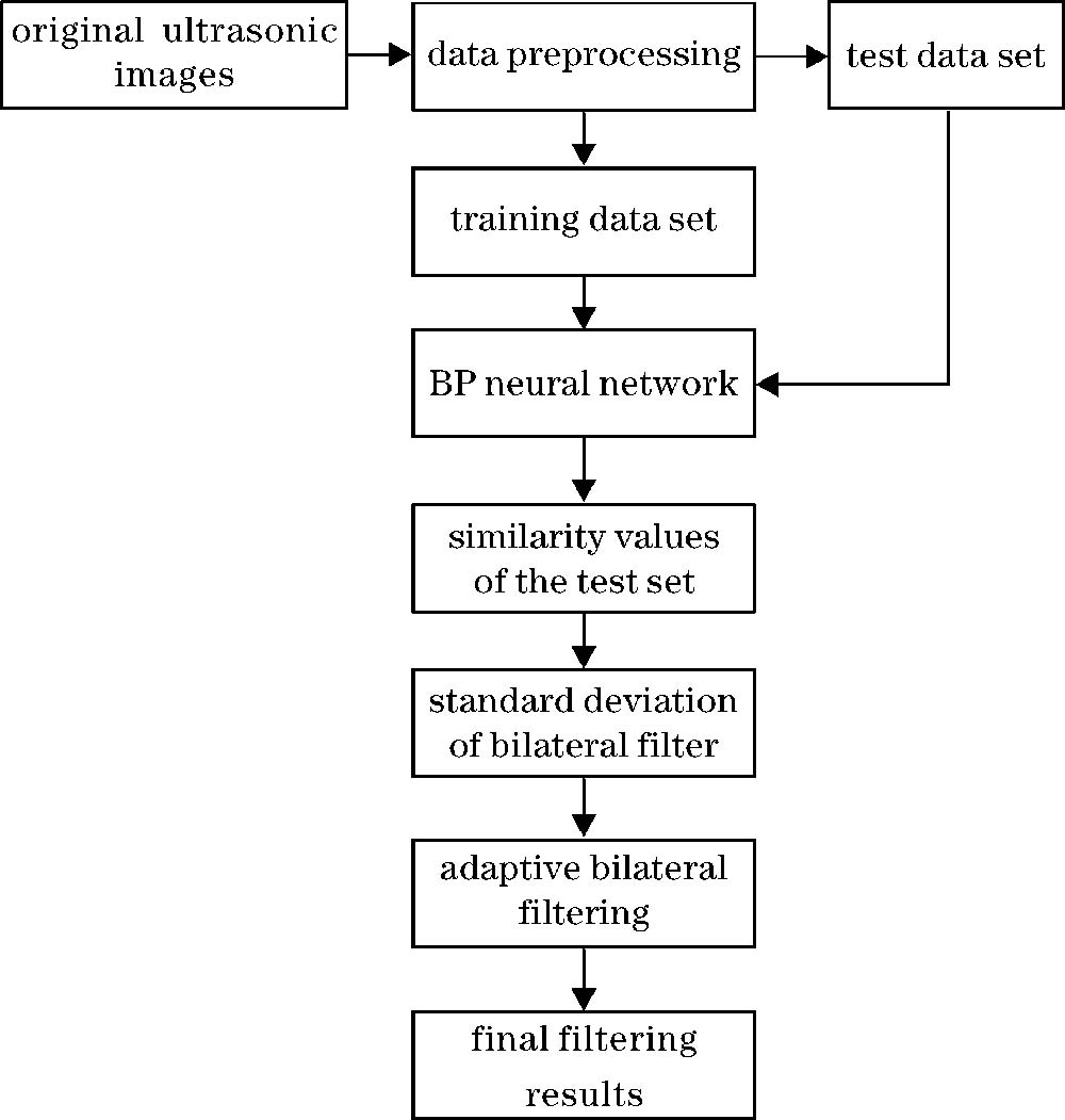
    Fig. 2. BP neural network based adaptive bilateral filtering model
    Denoising results of physical phantom ultrasonic image. (a) Original image; (b) P-M model; (c) DPAD method; (d) DnCNN model ; (e) our method
    Fig. 3. Denoising results of physical phantom ultrasonic image. (a) Original image; (b) P-M model; (c) DPAD method; (d) DnCNN model ; (e) our method
    Denoising results of liver ultrasonic image 1. (a) Original image; (b) P-M model; (c) DPAD method; (d) DnCNN model; (e) our method
    Fig. 4. Denoising results of liver ultrasonic image 1. (a) Original image; (b) P-M model; (c) DPAD method; (d) DnCNN model; (e) our method
    Denoising results of liver ultrasonic image 2. (a) Original image; (b) P-M model; (c) DPAD method; (d) DnCNN model; (e) our method
    Fig. 5. Denoising results of liver ultrasonic image 2. (a) Original image; (b) P-M model; (c) DPAD method; (d) DnCNN model; (e) our method
    Denoising results of kidney ultrasonic image. (a) Original image; (b) P-M model; (c) DPAD method; (d) DnCNN model; (e) our method
    Fig. 6. Denoising results of kidney ultrasonic image. (a) Original image; (b) P-M model; (c) DPAD method; (d) DnCNN model; (e) our method
    Image in figure 3Image size /(pixel×pixel)Window sizeIterationsSNR /dBCNR /dBupSNRupCNR
    (a)507×243----3.8423.951----
    (b)507×2433×310021.12820.367449.920415.490
    (c)507×2433×310027.42927.080613.930585.400
    (d)507×243----37.73736.843795.487778.818
    (e)507×2439×91033.26132.870765.720731.940
    Table 1. Objective analysis of denoising results of physical phantom ultrasonic image
    Image in figure 4Image size /(pixel×pixel)Window sizeIterationsSNR /dBCNR /dBupSNRupCNR
    (a)512×741----4.9054.492----
    (b)512×7413×310020.80219.383324.10331.50
    (c)512×7413×310019.02518.176287.87304.63
    (d)512×741----28.54027.089481.86503.05
    (e)512×7419×91025.71325.067424.22458.04
    Table 2. Objective analysis of denoising results of liver ultrasonic image 1
    Image in figure 5Image size /(pixel×pixel)Window sizeIterationsSNR /dBCNR /dBupSNRupCNR
    (a)715×901----5.4183.302----
    (b)715×9013×310018.47615.573241.00371.62
    (c)715×9013×310020.15717.652272.04434.59
    (d)715×901----24.23020.865347.21531.89
    (e)715×9019×91023.45819.069332.96477.50
    Table 3. Objective analysis of denoising results of liver ultrasonic image 2
    Image in figure 6Image size /(pixel×pixel)Window sizeIterationsSNR(dB)CNR(dB)upSNRupCNR
    (a)446×519----7.0136.676----
    (b)446×5193×310021.71320.148209.61201.80
    (c)446×5193×310022.07321.251214.74218.32
    (d)446×519----30.15428.563329.97327.85
    (e)446×5199×91028.73427.528309.72312.34
    Table 4. Objective analysis of denoising results of kidney ultrasonic image
    Xiaofang Zhu, Liang Jing, Dangguo Shao. Ultrasonic Image Denoising Using Adaptive Bilateral Filtering Based on Back Propagation Neural Network[J]. Laser & Optoelectronics Progress, 2020, 57(24): 241014
    Download Citation