• Acta Optica Sinica
  • Vol. 38, Issue 2, 0215005 (2018)
Yifeng Lu1、2, Qinhua Jin1, Jing Jing1, Yundai Chen1, Yihui Cao1, Jianan Li1, and Rui Zhu1、*
Author Affiliations
  • 1 Department of Cardiology, Chinese PLA General Hospital, Beijing 100853, China
  • 1 State Key Laboratory of Transient Optics and Photonics, Xi'an Institute of Optics and Precision Mechanics, Chinese Academy of Sciences, Xi'an, Shaanxi 710000, China
  • 2 University of Chinese Academy of Sciences, Beijing 100049, China
  • show less
    DOI: 10.3788/AOS201838.0215005 Cite this Article Set citation alerts
    Yifeng Lu, Qinhua Jin, Jing Jing, Yundai Chen, Yihui Cao, Jianan Li, Rui Zhu. Detection and Segmentation Algorithm for Bioresorbable Vascular Scaffolds Struts Based on Machine Learning[J]. Acta Optica Sinica, 2018, 38(2): 0215005 Copy Citation Text show less
    Workflow of BVS strut malapposition analysis (Local enlarged drawing in the first image shows structure of one of the BVS struts)
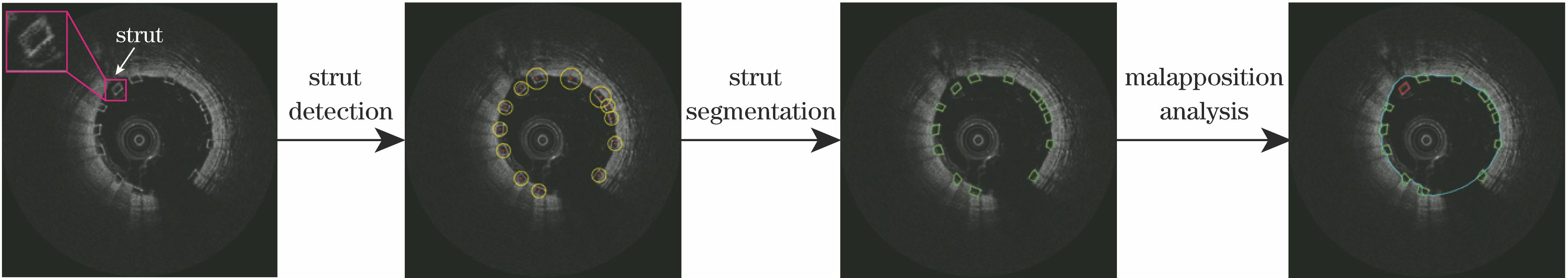
    Fig. 1. Workflow of BVS strut malapposition analysis (Local enlarged drawing in the first image shows structure of one of the BVS struts)
    (a) Single stump-based weak classifier; (b) strong classifier boosted by Fig. 2(a); (c) three-layer decision tree-based weak classifier; (d) strong classifier boosted by Fig. 2(c)
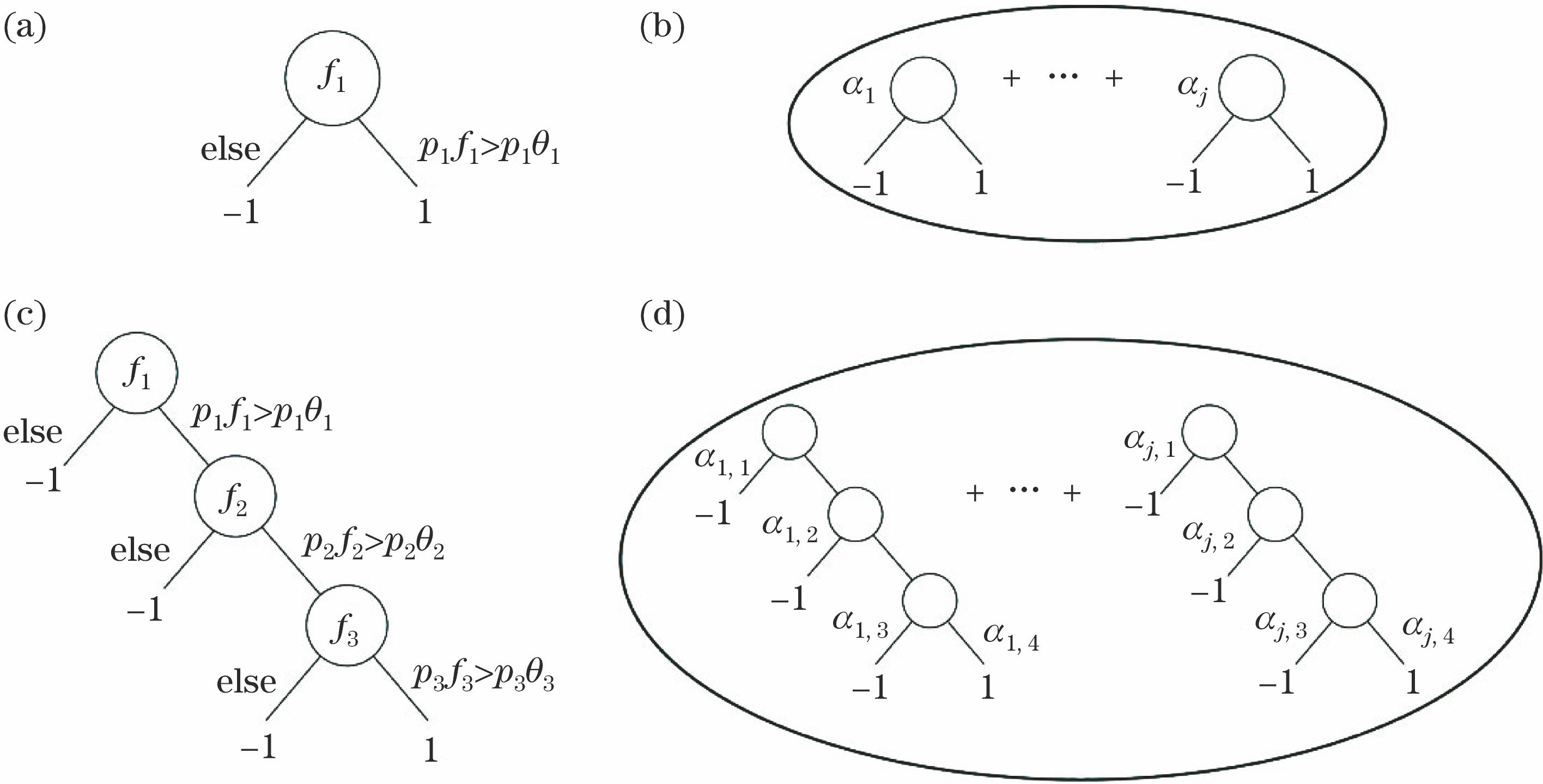
    Fig. 2. (a) Single stump-based weak classifier; (b) strong classifier boosted by Fig. 2(a); (c) three-layer decision tree-based weak classifier; (d) strong classifier boosted by Fig. 2(c)
    Structure of cascaded classifier
    Fig. 3. Structure of cascaded classifier
    Workflow of detection. (a) Input image; (b) detection region; (c) diagram of sliding sub-window; (d) detection through cascaded classifier; (e) BVS candidates; (f) output image
    Fig. 4. Workflow of detection. (a) Input image; (b) detection region; (c) diagram of sliding sub-window; (d) detection through cascaded classifier; (e) BVS candidates; (f) output image
    Procedure of strut segmentation. (a) Strut in Cartesian coordinate system; (b) strut in polar coordinate system; (c) segmented contour in polar coordinate system; (d) segmented contour transformed back into Cartesian coordinate system
    Fig. 5. Procedure of strut segmentation. (a) Strut in Cartesian coordinate system; (b) strut in polar coordinate system; (c) segmented contour in polar coordinate system; (d) segmented contour transformed back into Cartesian coordinate system
    Results of strut malapposition analysis. (a) Normal IVOCT images; (b)(c) images with blood artifacts; (d)-(f) images with both apposed and malapposed struts (For malapposed struts, distances between strut and lumen are represented by white lines)
    Fig. 6. Results of strut malapposition analysis. (a) Normal IVOCT images; (b)(c) images with blood artifacts; (d)-(f) images with both apposed and malapposed struts (For malapposed struts, distances between strut and lumen are represented by white lines)
    (a) Error curve of training; (b) ROC curve of testing
    Fig. 7. (a) Error curve of training; (b) ROC curve of testing
    Data setNumber of evaluated framesNumber of ground truthDetectionSegmentation
    Rre /%Rp /%FECP /cmCDice
    No.18169195.589.80.9326.40.80
    No.211992891.389.80.9131.20.79
    No.3118117292.089.40.9132.40.80
    No.47860490.788.40.9021.30.82
    No.5147118893.583.00.8824.80.82
    No.68663590.784.60.8630.00.80
    No.77660386.989.20.8824.10.79
    No.8150124092.183.20.8729.10.80
    Average--91.687.20.8927.40.80
    Table 1. Results of strut detection and segmentation
    Yifeng Lu, Qinhua Jin, Jing Jing, Yundai Chen, Yihui Cao, Jianan Li, Rui Zhu. Detection and Segmentation Algorithm for Bioresorbable Vascular Scaffolds Struts Based on Machine Learning[J]. Acta Optica Sinica, 2018, 38(2): 0215005
    Download Citation