• Acta Photonica Sinica
  • Vol. 51, Issue 6, 0618001 (2022)
Kaiqiu LAN1、2、*, Xibin YANG2, Baoteng XU2, Jialin LIU2, Wei ZHOU2, and Daxi XIONG2
Author Affiliations
  • 1School of Biomedical Engineering(Suzhou),Division of Life Sciences and Medicine,University of Science and Technology of China,Suzhou,Jiangsu 215163,China
  • 2Center of Light and Health,Suzhou Institute of Biomedical Engineering and Technology,Chinese Academy of Sciences,Suzhou,Jiangsu 215163,China
  • show less
    DOI: 10.3788/gzxb20225106.0618001 Cite this Article
    Kaiqiu LAN, Xibin YANG, Baoteng XU, Jialin LIU, Wei ZHOU, Daxi XIONG. In Vivo,Dual-color Fluorescent Imaging Miniature Microscope[J]. Acta Photonica Sinica, 2022, 51(6): 0618001 Copy Citation Text show less
    The cross-sectional view of DCFIMM-SBIand DCFIMM-DBI
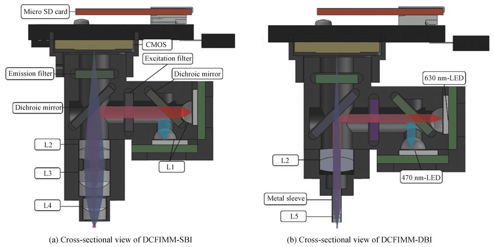
    Fig. 1. The cross-sectional view of DCFIMM-SBIand DCFIMM-DBI
    Schematic diagram of the lens focusing light of different wavelengths
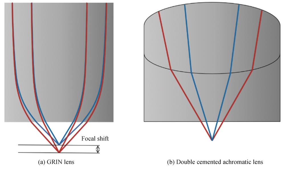
    Fig. 2. Schematic diagram of the lens focusing light of different wavelengths
    Optical path of optical system
    Fig. 3. Optical path of optical system
    Point diagram and chromatic focal shift of the optical systems
    Fig. 4. Point diagram and chromatic focal shift of the optical systems
    DCFIMM circuit principle
    Fig. 5. DCFIMM circuit principle
    Photograph of the DCFIMM imaging experiment
    Fig. 6. Photograph of the DCFIMM imaging experiment
    Results of the USAF resolution target imaging experiment
    Fig. 7. Results of the USAF resolution target imaging experiment
    Results of the fluorescent microsphere imaging experiment
    Fig. 8. Results of the fluorescent microsphere imaging experiment
    Results of the hybrid fluorescent microsphere imaging experiment
    Fig. 9. Results of the hybrid fluorescent microsphere imaging experiment
    Results of the hybrid fluorescent microsphere focusing and contrast imaging experiment
    Fig. 10. Results of the hybrid fluorescent microsphere focusing and contrast imaging experiment
    ComponentDimension/mmModel
    Outer casing22×17×143D printing(Wenext)
    470 nm-LED3×4.5×2LXML-PB01-0030(Lumileds)
    630 nm-LED3×4.5×2LXM2-PD01-0060(Lumileds)
    Dichroic mirror for combining excitation light5×6×1T495lpxr(Chroma)
    Excitation filter5×5×1ET-Fitc/Cy5x(Chroma)
    Dichroic mirror5×7×1TFITC/Cy5rpc(Chroma)
    Emission filter5×5×1ET-Fitc/Cy5m(Chroma)
    Collimator lens(L1)Diameter 347-269(Edmund)
    Imaging systemSee Table 2 for details
    PCB(including micro SD Card)17×23×4Custom design
    Battery14×10×6601215(Yuhuida)
    Metal sleeve1.2×4(thickness 0.1)Custom design
    Table 1. The main component of DCFIMM
    Imaging optical systemComponentDiameter /mmFocal length /mmModel
    L2DCFIMM-SBIImaging lens41263-692(Edmund)
    L3Objective lens4663-690(Edmund)
    L43645-089(Edmund)
    L2DCFIMM-DBIImaging lens41263-692(Edmund)
    L5Objective lens1365-566(Edmund)
    Table 2. The components of DCFIMM imaging optical systems
    Optical systemMagnificationNumerical aperture/(N.A.)Field of view/mm2Work distance/mm
    GRIN lens6.60.400.55×0.550.17
    DCFIMM-SBI3.20.411.10×1.100.60
    DCFIMM-DBI4.20.150.77×0.772.40
    Table 3. The main parameters of DCFIMM imaging optical systems
    Kaiqiu LAN, Xibin YANG, Baoteng XU, Jialin LIU, Wei ZHOU, Daxi XIONG. In Vivo,Dual-color Fluorescent Imaging Miniature Microscope[J]. Acta Photonica Sinica, 2022, 51(6): 0618001
    Download Citation