• Acta Optica Sinica
  • Vol. 38, Issue 5, 0517001 (2018)
Site Luo1, Yingwei Fan1, Wei Chang1, Xin An1、2, Hui Zhao1, and Li Huo1、*
Author Affiliations
  • 1 Department of Biomedical Engineering, Tsinghua University, Beijing 100084, China
  • 1 Department of Electronic Engineering, Tsinghua University, Beijing 100084, China
  • 1 Foshan Optomedic Technologies Co., Ltd., Foshan, Guangdong 528000, China
  • 1 Suzhou Institute of Nano-Tech and Nano-Bionics, Chinese Academy of Sciences, Suzhou, Jiangsu 215123, China
  • 2 Foshan Optomedic Technologies Co., Ltd., Foshan, Guangdong 528000, China
  • show less
    DOI: 10.3788/AOS201838.0517001 Cite this Article Set citation alerts
    Site Luo, Yingwei Fan, Wei Chang, Xin An, Hui Zhao, Li Huo. Boundary Region of Stomach Mucinous Carcinoma with Swept Source Optical Coherence Tomography[J]. Acta Optica Sinica, 2018, 38(5): 0517001 Copy Citation Text show less
    (a) OCT instrument; (b) tissue excised in stomach cancer operation
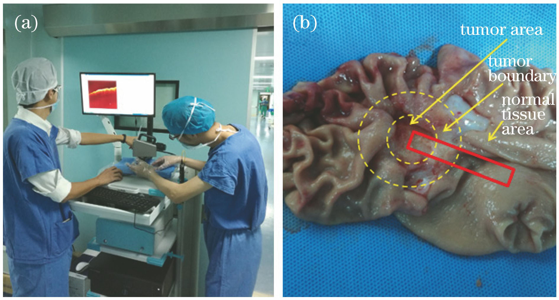
    Fig. 1. (a) OCT instrument; (b) tissue excised in stomach cancer operation
    (a) OCT image of normal stomach tissue of stomach cancerous patient; (b) corresponding pathological section
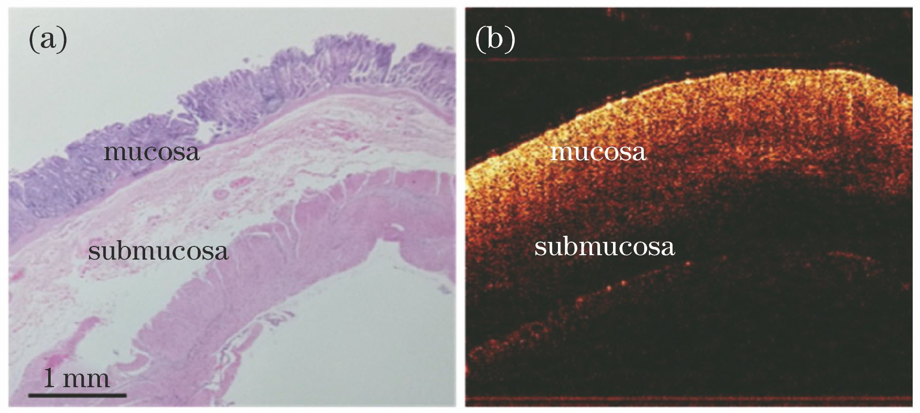
    Fig. 2. (a) OCT image of normal stomach tissue of stomach cancerous patient; (b) corresponding pathological section
    (a) Pathological section of stomach mucinous cancerous boundary region; (b)-(f) OCT images nearby corresponding pathological section
    Fig. 3. (a) Pathological section of stomach mucinous cancerous boundary region; (b)-(f) OCT images nearby corresponding pathological section
    (a) Stomach tissue volumetric image after 3D reconstruction; (b) stomach tissue volumetric image after surface flattening
    Fig. 4. (a) Stomach tissue volumetric image after 3D reconstruction; (b) stomach tissue volumetric image after surface flattening
    Layered images at different depths of stomach cancerous boundary region. (a) 25 pixels; (b) 35 pixels; (c) 45 pixels; (d) 55 pixels
    Fig. 5. Layered images at different depths of stomach cancerous boundary region. (a) 25 pixels; (b) 35 pixels; (c) 45 pixels; (d) 55 pixels
    Local standard deviation analysis of layered images at different depths of stomach cancerous boundary region. (a) 25 pixels; (b) 35 pixels; (c) 45 pixels; (d) 55 pixels
    Fig. 6. Local standard deviation analysis of layered images at different depths of stomach cancerous boundary region. (a) 25 pixels; (b) 35 pixels; (c) 45 pixels; (d) 55 pixels
    Site Luo, Yingwei Fan, Wei Chang, Xin An, Hui Zhao, Li Huo. Boundary Region of Stomach Mucinous Carcinoma with Swept Source Optical Coherence Tomography[J]. Acta Optica Sinica, 2018, 38(5): 0517001
    Download Citation