
Liu Lixin, Li Mengzhu, Zhao Zhigang, Qu Junle. Recent Advances of Hyperspectral Imaging Application in Biomedicine[J]. Chinese Journal of Lasers, 2018, 45(2): 207017
- Chinese Journal of Lasers
- Vol. 45, Issue 2, 207017 (2018)
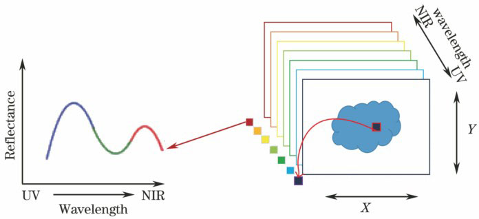
Fig. 1. 3D HSI images and spectra
![Schematic of a pushbroom HSI system[8]](/richHtml/zgjg/2018/45/2/0207017/img_2.jpg)
Fig. 2. Schematic of a pushbroom HSI system[8]
Fig. 3. Spectral reflectance of the normal mucosa and the tumor[10]
Fig. 4. Image processing by hyperspectral camera[10]. (a) Esophagogastroduodenoscopy image of a gastric adenoma of the anterior wall of the stomach; (b) hyperspectral camera image before image processing; (c) image processed on the basis of the SR at the 770 nm wavelength with a two-color gradation; (d) image processed on the basis of the SR at the 770 nm wavelength with a 20-color gradation by hyperspectral data analyzer
Fig. 5. Classification result of a pathology slide based on wide-gap second derivative analysis[14]. (a) The mean transmittance curves of two different regional organizations; (b) corresponding wide-gap SDT spectra; (c) the cancer tissue of the H&E-stained histological slide; (d) classification result according to wide-gap SDT method combined with segmentation algorithm
Fig. 6. In vivo rodent retinal snapshot HSI[32]. (a) Color fundus image recovered from 16 spectral channels; (b) representative spectra of the four labeled locations; (c) false-color fundus image with enhanced contrast between vessels and tissue
Fig. 7. Reflectance spectrum of pancreatic tissue[38]

Set citation alerts for the article
Please enter your email address
© Copyright 2018-2021 | Chinese Laser Press. All Rights Reserved 沪ICP备15018463号-20