Journals >Chinese Journal of Lasers
Contents


2018
Volume: 45 Issue 2
30 Article(s)
Export citation format
[in Chinese]

Introduction for Special Issue
[in Chinese], and [in Chinese]
Chinese Journal of Lasers
- Publication Date: Jan. 01, 1900
- Vol. 45, Issue 2, 207000 (2018)

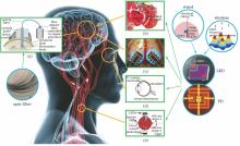
Implantable Optoelectronic Devices and Systems for Biomedical Application
Shi Zhao, Li Lizhu, Zhao Yu, Fu Ruxing, and Sheng Xing
Implantable electronic devices have become indispensable tools for biological science research and medical clinical applications. Focusing on the implantable optoelectronic devices and systems, we introduce the material fabrication, technique integration and implantable strategies of various implantable passive and actImplantable electronic devices have become indispensable tools for biological science research and medical clinical applications. Focusing on the implantable optoelectronic devices and systems, we introduce the material fabrication, technique integration and implantable strategies of various implantable passive and active devices for biomedical applications. The energy and information transmissions in implantable optoelectronic devices are classified. In addition, the examples of typical implantable electronic systems are showcased from the perspective of biomedical research and applications. Current methods and technologies are discussed comprehensively. In addition, the characteristics, future development trend and challenges of various technique schemes are analyzed and summarized when the requirements of practical applications and biocompatibility are considered..
Chinese Journal of Lasers
- Publication Date: Jan. 01, 1900
- Vol. 45, Issue 2, 207001 (2018)
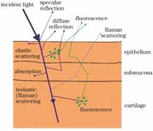
Real-Time in vivo Raman Spectroscopy and Its Clinical Applications in Early Cancer Detection
Shuang Wang, and Haishan Zeng
Raman spectroscopy (RS) is a unique analytical technique that probes molecular vibrations, and provides specific information about the structure and conformation of biomolecular constituents in biological tissues. Its capabilities for fast, accurate, and noninvasive/minimally invasive analysis have facilitated clinicalRaman spectroscopy (RS) is a unique analytical technique that probes molecular vibrations, and provides specific information about the structure and conformation of biomolecular constituents in biological tissues. Its capabilities for fast, accurate, and noninvasive/minimally invasive analysis have facilitated clinical applications in early cancer detection and pathophysiological investigations. Recent technology advancement in lasers, optical fiber probes and photoelectric devices has resulted in new rapid Raman spectroscopy systems with significantly better performance for real-time clinical measurements. Thereby, the scope and depth of its clinical usages have been expanded and deepened with the manifestation of its scientific connotation and diagnostic value. This short review provided an introduction to the theory and technology behind the development of integrated real-time Raman system for in vivo cancer detection. The perspective of its clinical utility, exemplified with skin and lung cancer detection, was presented with the intention of providing a useful reference for relevant basic research and technical innovation..
Chinese Journal of Lasers
- Publication Date: Jan. 01, 1900
- Vol. 45, Issue 2, 207002 (2018)
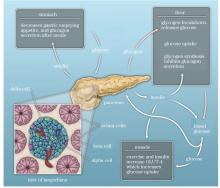
Research Advances in Blood Glucose Monitoring System
Sun Kai, Zhou Hua, Yang Yingkun, and Wu Changfeng
Diabetes mellitus is defined as a group of metabolic disorders characterized by hyperglycemia, which is one of the most serious health problems of the 21st century. Tight glycemic control is crucial for preventing diabetic complications. For more than 50 years of research, a considerable amount of glucose sensors is suDiabetes mellitus is defined as a group of metabolic disorders characterized by hyperglycemia, which is one of the most serious health problems of the 21st century. Tight glycemic control is crucial for preventing diabetic complications. For more than 50 years of research, a considerable amount of glucose sensors is successively developed, and different innovative detection strategies are constantly developed. For blood glucose monitoring system, we review the history, expound the principles and advantages, discuss the recent developments and current status, and outline key challenges and opportunities in the future..
Chinese Journal of Lasers
- Publication Date: Jan. 01, 1900
- Vol. 45, Issue 2, 207003 (2018)
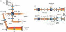
Research Progress on Parallel Spectral Domain Optical Coherence Tomography Technology
Shen Yi, Chen Zhiyan, Qiu Jianrong, and Ding Zhihua
Parallel spectral domain optical coherence tomography (PSD-OCT) technology is introduced. Research progresses on the method and system development of PSD-OCT in Zhihua Ding′s group are mainly reported, which includes the wild-field PSD-OCT system based on global optimization, the discrimination method for real and fakeParallel spectral domain optical coherence tomography (PSD-OCT) technology is introduced. Research progresses on the method and system development of PSD-OCT in Zhihua Ding′s group are mainly reported, which includes the wild-field PSD-OCT system based on global optimization, the discrimination method for real and fake defects based on the complex interference signal and phase information, and the approach for correcting motion artifacts based on the spatial-spectral encoding and overlapped data correlation algorithm. The established PSD-OCT system can be used to identify the surface and internal defects of glass, which avoids the misjudgment of fake defects. With the introduction of spatial-spectral encoding units, this system can also be used to correct uncontrolled motions..
Chinese Journal of Lasers
- Publication Date: Jan. 01, 1900
- Vol. 45, Issue 2, 207004 (2018)

Viscoelasticity Measurement of Biological Tissues Using Laser Speckle Techniques: a Review
Chen Xiao, Lu Jinling, and Li Pengcheng
Viscosity and elasticity of biological tissues will change during onset and progression of many diseases. Viscoelasticity measurement of biological tissues plays an important role in diagnosis of diseases. We particularly review some basic principles and recent advances of viscoelasticity measurement based on laser speViscosity and elasticity of biological tissues will change during onset and progression of many diseases. Viscoelasticity measurement of biological tissues plays an important role in diagnosis of diseases. We particularly review some basic principles and recent advances of viscoelasticity measurement based on laser speckle techniques from three aspects: difference of laser speckle contrast under elastic waves, optical intensity autocorrelation curve under Brownian motion, and laser speckle shift under low frequency oscillation stress..
Chinese Journal of Lasers
- Publication Date: Jan. 01, 1900
- Vol. 45, Issue 2, 207005 (2018)
Laser Speckle Contrast Imaging on in vivo Blood Flow: a Review
Li Chenxi, Chen Wenliang, Jiang Jingying, Fan Ying, Yang Jingzi, and Xu Kexin
Based on the dynamic light scattering and approximate model, laser speckle contrast imaging (LSCI) is a powerful tool for full-field imaging of blood flow by analyzing the characteristics of speckle space or time intensity fluctuation. Because of the advantages of large imaging area, fast speed and high resolution, theBased on the dynamic light scattering and approximate model, laser speckle contrast imaging (LSCI) is a powerful tool for full-field imaging of blood flow by analyzing the characteristics of speckle space or time intensity fluctuation. Because of the advantages of large imaging area, fast speed and high resolution, the technology is used in biomedical imaging research and clinical diagnosis widely. A lot of research on the theoretical models, imaging methods and applications of LSCI are done by many researchers. This review presents the main improvement on method and applications of LSCI. Moreover, we discuss the concepts about increasing resolution, contrast, imaging penetration, and quantitative ability of LSCI. Finally, we review the applications of LSCI in ophthalmology, microcirculation, neuroscience, dermatology and surgical monitoring..
Chinese Journal of Lasers
- Publication Date: Jan. 01, 1900
- Vol. 45, Issue 2, 207006 (2018)
Tumor-Microenvironment Activable Smart Nanocarrier System for Photodynamic Therapy of Cancers
Lan Shanyou, Zhang Da, Liu Xiaolong, and Zeng Yongyi
Photodynamic therapy is a noninvasive therapy of cancers that utilizes exogenous visible, near-infrared light to induce the photosensitizer to react with molecular oxygen to produce a highly active photochemical product of reactive oxygen species which can induce apoptosis or necrosis of tumor cells, with low immunogenPhotodynamic therapy is a noninvasive therapy of cancers that utilizes exogenous visible, near-infrared light to induce the photosensitizer to react with molecular oxygen to produce a highly active photochemical product of reactive oxygen species which can induce apoptosis or necrosis of tumor cells, with low immunogenicity, low cost, high selectivity and other characteristics. Photodynamic therapy has become a hot topic in the basic research and clinical transformation of cancer treatment. However, the decreased effect of photodynamic therapy induced by tumor-microenvironment factors and the tolerance of tumor cells hinder the development of photodynamic therapy and clinical application. With the rapid development of comprehensive fields such as nanotechnology and biomedical engineering, and the deepening study of tumor microenvironment, the tumor-microenvironment activable smart nanocarrier system for the diagnosis and the synergistic treatment of cancer draw high attention in recent years. Based on this, the research of tumor-microenvironment activable smart nanocarrier system is reviewed to provide a reference and a new research idea for photodynamic therapy of tumors..
Chinese Journal of Lasers
- Publication Date: Jan. 01, 1900
- Vol. 45, Issue 2, 207008 (2018)
Progress on Methods of Quantitative Phase Measurement and Retrieval for Biological Cells
Zhang Lu, Zhao Chunhui, Kang Senbai, Zhao Hong, Zhang Chunwei, and Yuan Li
The phase measurement methods for label-free cells, as non-invasive methods, are applied to realizing the quantitative reconstruction for spatial morphologies of static and dynamic biological samples. It can provide implementable conditions for visualizing complex biophysical information in the process of cell dynamic The phase measurement methods for label-free cells, as non-invasive methods, are applied to realizing the quantitative reconstruction for spatial morphologies of static and dynamic biological samples. It can provide implementable conditions for visualizing complex biophysical information in the process of cell dynamic detection. The novel phase detection technologies for dynamic cells are mainly described, such as the synchronous phase shift methods, the digital holographic methods, and the methods for focused fluid. The traditional phase detection technologies for static cells are reviewed briefly. Then the key parameters of these technologies are compared, such as sampling rate, imaging resolution, and detection accuracy. Furthermore, we illustrate their application fields considering their various biological information detections. Finally, the basic principles, technical characteristics and recent developments for various phase retrieval technologies are analyzed and summarized..
Chinese Journal of Lasers
- Publication Date: Jan. 01, 1900
- Vol. 45, Issue 2, 207009 (2018)
Applications of Two-Photon Excitation Fluorescence Lifetime Imaging in Tumor Diagnosis
Li Hui, Xia Xianyuan, Chen Tingai, Yu Jia, Li Xi, and Zheng Wei
Two-photon excitation fluorescence lifetime imaging is an ingenious technique which can provide both morphological and biochemical information of tissues in three-dimensional space with subcellular resolution. The structural and functional properties of tissue can be quantitatively characterized in detail. Therefore, tTwo-photon excitation fluorescence lifetime imaging is an ingenious technique which can provide both morphological and biochemical information of tissues in three-dimensional space with subcellular resolution. The structural and functional properties of tissue can be quantitatively characterized in detail. Therefore, this technique provides a powerful tool for the noninvasive, label-free, intravital imaging of biological tissues, and which is promising to be applied in clinical diagnosis of tumors. In recent years, this technology has been used to detect tumors, and which has already become one of the research foci in the field of biomedicine. In this review paper, firstly, the concept of the two-photon excitation fluorescence lifetime and common fluorescence lifetime detection methods are introduced simply; secondly, we summarize the recent progresses of using two-photon excitation fluorescence lifetime imaging to detect various tumors, including digestive tract tumors, brain tumors, skin tumors, and so on, and among them, our results on the detection of gastric cancers and glioma are elaborately depicted; Finally, the potential advantages and possible challenges of this technology in future clinical application are provided and discussed..
Chinese Journal of Lasers
- Publication Date: Jan. 01, 1900
- Vol. 45, Issue 2, 207010 (2018)
Research Progress in Fourier Domain Optical Coherence Tomography
Li Pei, Yang Shanshan, Ding Zhihua, and Li Peng
Optical coherence tomography (OCT) enables three-dimensional internal micro-structural imaging of biological tissue in real time with high resolution, and it has wide range of applications in clinic diagnosis, treatment and basic scientific research. In recent years, thanks to the development of light sources and detecOptical coherence tomography (OCT) enables three-dimensional internal micro-structural imaging of biological tissue in real time with high resolution, and it has wide range of applications in clinic diagnosis, treatment and basic scientific research. In recent years, thanks to the development of light sources and detection technology, Fourier domain OCT has become the mainstream OCT model, particularly has promoted the development of functional imaging technology, such as OCT microangiography. In this work, we review the principle of OCT based on Fourier domain OCT, and illustrate the primary performance parameters and their influence factors of system. The recent advances and expectations in imaging range, speed and functional extensions are introduced..
Chinese Journal of Lasers
- Publication Date: Jan. 01, 1900
- Vol. 45, Issue 2, 207011 (2018)
Progress in Biomagnetic Signal Measurements with Ultra-Sensitive Atomic Magnetometers
Wang Xiaofei, Sun Xianping, Zhao Xiuchao, Zhu Maohua, Ye Chaohui, and Zhou Xin
Biomagnetic fields produced by organisms carry valuable informations of electrophysiologies and pathologies, which are commonly detected by superconducting quantum interference device. As an ultra-sensitive magnetic field detector with the sensitivity of femtotesla level, the ultra-sensitive atomic magnetometer plays aBiomagnetic fields produced by organisms carry valuable informations of electrophysiologies and pathologies, which are commonly detected by superconducting quantum interference device. As an ultra-sensitive magnetic field detector with the sensitivity of femtotesla level, the ultra-sensitive atomic magnetometer plays an important role in detection and study of biomagnetism. We introduce the sources and characteristics of biological magnetic field signals, as well as the physical mechanism and classification of ultra-sensitive atomic magnetometers. We also summarize and forecast the applications of ultra-sensitive atomic magnetometers in the field of the biomagnetic field detection, such as magnetocardiography, magnetoencephalography, and neurosciences..
Chinese Journal of Lasers
- Publication Date: Jan. 01, 1900
- Vol. 45, Issue 2, 207012 (2018)
Research Progress on Photodynamic Antimicrobial Chemotherapy Based on Rare Earth Upconversion Nanoplatform
Zhao Yiming, Liu Chengcheng, Wang Jing, and Hu Min
Rare earth upconversion nanoparticles (UCNPs) can transform near-infrared light into ultraviolet-visible light, which can effectively solve the problems such as a low tissue penetration depth and a low treatment efficiency of photodynamic antimicrobial chemotherapy (PACT). The research progress on the photodynamic antiRare earth upconversion nanoparticles (UCNPs) can transform near-infrared light into ultraviolet-visible light, which can effectively solve the problems such as a low tissue penetration depth and a low treatment efficiency of photodynamic antimicrobial chemotherapy (PACT). The research progress on the photodynamic antimicrobial chemotherapy based on rare earth upconversion nanoplatforms is reviewed. The development of novel co-antibacterial platform and its clinical applications are prospected..
Chinese Journal of Lasers
- Publication Date: Jan. 01, 1900
- Vol. 45, Issue 2, 207013 (2018)
Application of Multiphoton Microscopy in Disease Diagnosis
Lin Hongxin, Zuo Ning, Zhuo Shuangmu, and Chen Jianxin
Multiphoton microscopy (MPM) technology offers non-destructive, non-labeled imaging of tissue by detecting optical signals such as two-photon excitation fluorescence and second harmonic produced by the interaction of femtosecond laser with internal components of biological tissue. MPM has the advantages of tissue microMultiphoton microscopy (MPM) technology offers non-destructive, non-labeled imaging of tissue by detecting optical signals such as two-photon excitation fluorescence and second harmonic produced by the interaction of femtosecond laser with internal components of biological tissue. MPM has the advantages of tissue microstructure sensitivity, high spatial resolution imaging, low killing of biological tissues, deep imaging depth, and the ability to obtain the biochemical information of the tissue. It has great potential in disease diagnosis. The review mainly focuses on the principle of MPM and its applications in disease diagnosis of gastrointestinal cancer, skin diseases and corneal diseases, and looks forward to the development of MPM..
Chinese Journal of Lasers
- Publication Date: Jan. 01, 1900
- Vol. 45, Issue 2, 207014 (2018)
Breath Analysis Using Laser Spectroscopy Techniques: Development and Future
Jiang Chenyu, Sun Meixiu, Li Yingxin, and Wang Chuji
As a promising new field of medicine and a new development direction of medical instrumentation, breath analysis offers noninvasive and real-time disease diagnostics and metabolic status monitoring potentially. Numerous breath biomarkers are detected and quantified so far by the utilization of gas chromatography-mass sAs a promising new field of medicine and a new development direction of medical instrumentation, breath analysis offers noninvasive and real-time disease diagnostics and metabolic status monitoring potentially. Numerous breath biomarkers are detected and quantified so far by the utilization of gas chromatography-mass spectrum(GC-MS) technique. Recent advances in laser spectroscopic techniques and laser sources accelerate the development of breath analysis. Compared with the MS-based techniques, laser spectroscopic techniques have not only the high-sensitivity and high-selectivity, but also the advantageous features of real-time response, low instrument cost and point-of-care testing(POCT). In the approximately 35 established breath biomarkers, serious species in human breath are analyzed by laser spectroscopic techniques, namely, tunable diode laser absorption spectroscopy (TDLAS), cavity ringdown spectroscopy (CRDS), and photoacoustic spectroscopy (PAS). The spectral fingerprints of the measured biomarkers are spanned from the ultra violet to the mid-infrared spectral regions..
Chinese Journal of Lasers
- Publication Date: Jan. 01, 1900
- Vol. 45, Issue 2, 207015 (2018)
Super-Resolution Localization Microscopy with Scientific Complementary Metal Oxide Semiconductor Camera
Gui Dan, Shang Mingtao, and Huang Zhenli
Super-resolution localization microscopy (SRLM) is a representative super-resolution imaging technology. Low-light detector is an essential component in SRLM. Compared with traditional electron multiplying charge coupled device (EMCCD) cameras using serial output, scientific complementary metal oxide semiconductor (sCMSuper-resolution localization microscopy (SRLM) is a representative super-resolution imaging technology. Low-light detector is an essential component in SRLM. Compared with traditional electron multiplying charge coupled device (EMCCD) cameras using serial output, scientific complementary metal oxide semiconductor (sCMOS) cameras with parallel output provide simultaneously large field of view, high imaging speed and low read noise, thus bring new opportunities for SRLM, especially on achieving video-rate imaging and large field of view imaging. At the same time, SRLM based on sCMOS camera is also facing several challenges which are mainly originated from the high-speed camera, including but not limited to massive data transmission, storage, and analysis. This paper introduces the principle and development of SRLM, and discusses the current status, opportunities and challenges of SRLM based on sCMOS camera..
Chinese Journal of Lasers
- Publication Date: Jan. 01, 1900
- Vol. 45, Issue 2, 207016 (2018)
Recent Advances of Hyperspectral Imaging Application in Biomedicine
Liu Lixin, Li Mengzhu, Zhao Zhigang, and Qu Junle
Hyperspectral imaging (HSI) combines spectroscopy and imaging technology, and can acquire multi-dimensional information that includes two-dimensional spatial information and one-dimensional spectral information. The space-resolved spectral images obtained by HSI can provide diagnostic information about histophysiology,Hyperspectral imaging (HSI) combines spectroscopy and imaging technology, and can acquire multi-dimensional information that includes two-dimensional spatial information and one-dimensional spectral information. The space-resolved spectral images obtained by HSI can provide diagnostic information about histophysiology, morphology, and composition of tissues. HSI is an emerging medical imaging modality and has broad potential applications in the field of biomedicine, especially in disease diagnosis and image-guided surgery. We introduce the basic principles of HSI, the configuration and characteristics of HSI system, and review the recent developments of HSI in biomedical field and its applications in disease diagnosis and surgical guidance. The future research direction and development prospects of HSI are also analyzed and forecasted..
Chinese Journal of Lasers
- Publication Date: Jan. 01, 1900
- Vol. 45, Issue 2, 207017 (2018)
Application Progress of Lensless Microscopy Imaging Technology for Point-of-Care Testing
Li Conghui, Cao Ruofan, Xu Xiayu, Li Fei, Zhang Zhenxi, and Xu Feng
Microorganisms, such as bacteria, are always detection objects in point-of-care testing, such as infectious diseases diagnosis, food safety monitoring and environment pollution detection. Optical microscopy is always a tool which is used to detect and analyze these detection objects. Lensless microscopy combines the saMicroorganisms, such as bacteria, are always detection objects in point-of-care testing, such as infectious diseases diagnosis, food safety monitoring and environment pollution detection. Optical microscopy is always a tool which is used to detect and analyze these detection objects. Lensless microscopy combines the sample with photodetectors such as charge coupled device (CCD) or complementary metal-oxide semiconductor (CMOS). This technique does not need optical devices, and the samples are imaged directly. The device is significantly simpler, smaller, easier to manipulate and cheaper than that of lens-based conventional microscope, which has been used in microstructure examinations, cell morphological analysis and microorganism detection. The lensless microscopic technology can be divided into three types, such as shadow imaging, fluorescence imaging and digital inline holography according to imaging principle. This paper reviews imaging principle and physical structure of three kinds of lensless microscopic technologies, and summaries the applications of lensless microscopic technologies in point-of-care testing. At last, we discuss the future development of lensless microscopic technologies..
Chinese Journal of Lasers
- Publication Date: Jan. 01, 1900
- Vol. 45, Issue 2, 207018 (2018)
Research Progress on Optical Coherence Tomography in Detecting Vascular Flow Field
Gao Feng, Fan Jinyu, Kong Wen, and Shi Guohua
Optical coherence tomography (OCT) can image the structure of the biological samples with high resolution and non-destruction. By analyzing the phase or intensity change of the signal, we can distinguish the motion tissue and the static tissue in the sampler, and then extract flow fieldfield information. Various methodOptical coherence tomography (OCT) can image the structure of the biological samples with high resolution and non-destruction. By analyzing the phase or intensity change of the signal, we can distinguish the motion tissue and the static tissue in the sampler, and then extract flow fieldfield information. Various methods are summarized to detect blood flow in blood vessel. OCT has become a powerful tool for the clinical measurement of vascular microcirculation, since it can image blood vessel in skin or retina without any auxiliary reagent. We reviewed six typical methods in Fourier OCT: phase-resolved Doppler OCT, phase-resolved Doppler variance, intensity-based Doppler variance, speckle variance, split-spectrum amplitude-decorrelation angiography, and optical micro-angiography. Each method has its own advantages and disadvantages on phase stabilization, flow rate measurement, signal-to-noise ratio, and real time. Imaging schemes are optimized based on the six methods. Finally, the prospects of OCT are included..
Chinese Journal of Lasers
- Publication Date: Jan. 01, 1900
- Vol. 45, Issue 2, 207019 (2018)
Progress in Organic Nanomaterials for Laser-Induced Photothermal Therapy of Tumor
Liang Guohai, and Xing Da
Laser-induced thermal therapy, which has many advantages including rapid operation, minimal damage and insignificant toxicity and side effects, is considered to be a promising non-invasive/minimally invasive therapeutic methodology, and it attracts much attention in recent years. In order to improve the therapeutic effLaser-induced thermal therapy, which has many advantages including rapid operation, minimal damage and insignificant toxicity and side effects, is considered to be a promising non-invasive/minimally invasive therapeutic methodology, and it attracts much attention in recent years. In order to improve the therapeutic effects, researchers study various types of photothermal therapy agents (PTAs). Compared with inorganic nanomaterials, PTAs based on organic nanomaterials have superior biocompatibility and biodegradability, and they are more promising for clinical translation. As a result, PTAs based on organic nanomaterials has rapid development in recent years. In this review, the recent progress of several typical classes of organic photothermal nano-agents, including near-infrared dye-containing micelles, protein-based photothermal agents, porphysomes, and conjugated polymers, is introduced, and their individual advantages and shortcomings are introduced. At last, the challenges and future direction of this field also be discussed..
Chinese Journal of Lasers
- Publication Date: Jan. 01, 1900
- Vol. 45, Issue 2, 207020 (2018)
Application of Gold-Nanoparticle-Based Fluorescent Molecular Beacons in BRCA1 mRNA Detection
Liu Ruonan, Li Tingting, Zhou Qiumei, and Gu Yueqing
In order to detect the expression of messenger ribonucleic acid (mRNA) of the breast cancer susceptibility gene 1 (BRCA1) ) in real time in living cells, a novel gold-nanoparticle-based fluorescent molecular beacon is designed and successfully prepared. The specificity, sensitivity, stability and toxicity of this beacoIn order to detect the expression of messenger ribonucleic acid (mRNA) of the breast cancer susceptibility gene 1 (BRCA1) ) in real time in living cells, a novel gold-nanoparticle-based fluorescent molecular beacon is designed and successfully prepared. The specificity, sensitivity, stability and toxicity of this beacon are investigated, the fluorescence recovery strength of this beacon is detected by the techniques of laser confocal microscopy and flow cytometry, and thus the expression of BRCA1 mRNA in human gastric cancer cell BGC823 and its drug-resistant cell BGC823/DDP is realized. The reverse transcription polymerase chain reaction (RT-PCR) technique is used to verify the reliability of this beacon for detecting the expression of BRCA1 mRNA in cells. The results show that this beacon has excellent characteristics and can be reliably used to detect the expression level of BRCA1 mRNA in tumor cells..
Chinese Journal of Lasers
- Publication Date: Jan. 01, 1900
- Vol. 45, Issue 2, 207021 (2018)
Wavelength Misalignment Analysis and Spectral Calibration for Fourier Domain Polarization-Sensitive Optical Coherence Tomography
Chen Yan, Li Zhongliang, Nan Nan, Bu Yang, Lu Yu, Song Siyu, and Wang Xiangzhao
Fourier domain polarization-sensitive optical coherence tomography (FD-PS-OCT) can measure the polarization properties of a sample to provide early diagnosis for some diseases. The accurate diagnosis is required to measure the polarization parameters in a high accuracy. However, the accuracy is greatly affected by waveFourier domain polarization-sensitive optical coherence tomography (FD-PS-OCT) can measure the polarization properties of a sample to provide early diagnosis for some diseases. The accurate diagnosis is required to measure the polarization parameters in a high accuracy. However, the accuracy is greatly affected by wavelength misalignment. Even a slight wavelength mismatch in FD-PS-OCT will result in nonnegligible polarization artifacts. So an accurate wavelength assignment is necessary. In order to decrease the polarization error and improve the accuracy of spectral calibration, the effect of the wavelength misalignment on the polarization parameters calculation should be clear. An analysis is performed on the relationship between the wavenumber misalignment and the calculated error in theory and simulation. Based on the error analysis, a spectral calibration method is proposed. The method achieves the wavelength alignment through evaluating the retardation and fast axis orientation errors. The wave plate experiments show the good performance of the method and the reasonability of the error analysis..
Chinese Journal of Lasers
- Publication Date: Jan. 01, 1900
- Vol. 45, Issue 2, 207022 (2018)
Growth Stages Characteristic Parameters of Bacteria in Water Based on Multi-Wavelength Transmission Spectra
Hu Yuxia, Zhao Nanjing, Gan Tingting, Duan Jingbo, Liu Jianguo, and Liu Wenqing
Research on the laws of bacterial growth and rapid acquisition of the changes in concentration, structure and chemical composition of bacterial cells can provide evidence for fast bacterial detection in environmental assessment and microbiological research fields. The transmission spectra in the region of 240-900 nm ofResearch on the laws of bacterial growth and rapid acquisition of the changes in concentration, structure and chemical composition of bacterial cells can provide evidence for fast bacterial detection in environmental assessment and microbiological research fields. The transmission spectra in the region of 240-900 nm of Escherichia coli suspension during growth are recorded by ultraviolet-visible spectrophotometry. The measured transmission spectra in the visible regions are interpreted based on Mie scattering theory, and the information of concentration and size of bacteria during growth are acquired. According to the ultraviolet absorption spectra of bacteria, the nucleic acid content is calculated with the absorption characteristics of chromophore amino acid and nucleic acid. The results show that the parameters interpreted from the spectra can reflect changes in concentration, size and chemical composition of bacteria during growth. These changes demonstrate that Escherichia coli cells undergo different growth stages under suitable growth condition, which is consistent with the laws of bacterial growth. Multi-wavelength transmission spectroscopy can realize bacteria motion-tracking detection during growth, and can obtain morphological characteristics and chemical composition of bacteria, which provides a new method for multiple parameters measurement of bacteria..
Chinese Journal of Lasers
- Publication Date: Jan. 01, 1900
- Vol. 45, Issue 2, 207023 (2018)
Study of Stripe Regeneration of Zebrafish Based on Laser Ablation Technology
Jiang Pengchong, Wei Wei, Hu Fen, Zhang Xinzheng, and Pan Leiting
In the present work, 355 nm nanosecond pulsed laser is used to ablate zebrafish surface stripes to investigate the mechanism underlying formation and regeneration of pigment cells pattern. The experimental results show that zebrafish surface xanthophore clusters can only promote melanophore regeneration within the distIn the present work, 355 nm nanosecond pulsed laser is used to ablate zebrafish surface stripes to investigate the mechanism underlying formation and regeneration of pigment cells pattern. The experimental results show that zebrafish surface xanthophore clusters can only promote melanophore regeneration within the distance of half width of a stripe but not close to each other. Meanwhile, xanthophores significantly affect melanophore regeneration in a direction perpendicular to its stripe rather than parallel to its stripe, indicating the orientation specificity. The results can provide new experimental supports for understanding how zebrafish self-organize spatially periodic patterns..
Chinese Journal of Lasers
- Publication Date: Jan. 01, 1900
- Vol. 45, Issue 2, 207024 (2018)
Simulation Study on Methodological Feasibility of Diffuse Raman Tomography
Wu Yutong, Jia Mengyu, Wang Bingyuan, Zhao Huijuan, and Gao Feng
Diffuse Raman tomography (DRT) is a new in vivo, label-free, three-dimensional optical imaging technique, and it combines the molecular fingerprint of Raman spectrum and the depth resolution of diffuse optical tomography. A feasibility study on the DRT method based on the Monte-Carlo photon migration model is proposed.Diffuse Raman tomography (DRT) is a new in vivo, label-free, three-dimensional optical imaging technique, and it combines the molecular fingerprint of Raman spectrum and the depth resolution of diffuse optical tomography. A feasibility study on the DRT method based on the Monte-Carlo photon migration model is proposed. The proposed method establishes a fast and effective Raman spectra data simulator according to the Green′s reciprocity theorem,which can objectively simulate the measured Raman spectra data at different realistic signal-to-noise ratios. Combined with the linear reconstruction technique of diffuse fluorescence tomography, the proposed method can be used in the multi-wavelength Raman spectral analysis of laminar tissues. A laminar tissue model composed of five biochemical compositions is simulated and reconstructed from 178 wavelength Raman spectral analysis, which shows that linear reconstruction technique of the proposed DRT can construct effectively the three-dimensional relative concentration distributions of different components..
Chinese Journal of Lasers
- Publication Date: Jan. 01, 1900
- Vol. 45, Issue 2, 207025 (2018)
Study on Photoacoustic Effect in Nanoscale and Photoacoustic Conversion Mechanism of Nanoprobes
Shi Yujiao, and Xing Da
The construction of nanoprobes with high conversion efficiency is the key factor for the development of photoacoustic (PA) molecular imaging. The traditional design methods for PA probes usually maximize their optical absorption at the tissue optical window, while the research on the PA conversion properties of the proThe construction of nanoprobes with high conversion efficiency is the key factor for the development of photoacoustic (PA) molecular imaging. The traditional design methods for PA probes usually maximize their optical absorption at the tissue optical window, while the research on the PA conversion properties of the probes has not been given enough attention. Taking gold nanospheres as an example, the mechanism of microcosmic PA conversion of nanoprobes in PA effect mediated by thermal expansion mechanism is discussed, which makes the rational design of probe with high conversion efficiency possible. Based on theoretical analysis and finite element analysis, it is indicated that nanoprobes no longer satisfy the thermal confinement conditions due to the small size effect. The thermal energy diffuses from the sphere to the surrounding medium in the range of laser pulse. As a result, the PA signal is composed of nanoprobes itself and the thermal expansion of media around the nanoprobes..
Chinese Journal of Lasers
- Publication Date: Jan. 01, 1900
- Vol. 45, Issue 2, 207026 (2018)
Laser Thermal Damage Evaluation of Biological Tissues Based on Monitoring of Dual-Point Optical Parameters
Jia Weiwei, Dai Lijuan, Hua Guoran, and Qian Zhiyu
The scattering intensity at 720 nm (I720) is chosen as the optical parameter of evaluation,and we use the dual point optical parameters far from the center to evaluate the thermal damage near the center during the process of laser thermal damage. Firstly, we test on the laser thermal damage of in vitro porcine livers tThe scattering intensity at 720 nm (I720) is chosen as the optical parameter of evaluation,and we use the dual point optical parameters far from the center to evaluate the thermal damage near the center during the process of laser thermal damage. Firstly, we test on the laser thermal damage of in vitro porcine livers to collect I720 in real time at points which are 4, 8, 12 mm far from the heating center, and laser heating is stopped when I720 at 8 mm rises to 6 times compared with its initial value (group A). We conduct twenty experiments to calculate rising multiple of I720 at 4 mm far from the center with the mathematical model using the parameters of heating time, rising multiples of I720 at 8 mm and 12 mm. Secondly, we repeat the above thermal damage experiments and real-time data acquisition when the condition of stopping heating is I720 at 4 mm rising to 3, 4, 5, 6 times (group B). Ten experiments are done for each condition, and forty experiments are established. Finally, the slice analysis on samples of group B are taken to establish the relationship between the rising multiples of I720 and the damage effect. The results show that the accuracy is above 90% when heating time and rising multiples of I720 at 8 mm and 12 mm are used to estimate rising multiple of I720 at 4 mm. It is feasible to evaluate the effect of laser thermal damage in real time at a point near the damage center by dual-point optical parameters far from the center..
Chinese Journal of Lasers
- Publication Date: Jan. 01, 1900
- Vol. 45, Issue 2, 207027 (2018)
Detection of Antibiotics Based on Hyphenated Technique of Electrostatic-Preconcentration and Surface-Enhanced-Raman-Spectroscopy
Ma Haikuan, Zhang Xu, Zhong Shilei, Shi Xiaofeng, Ma Lizhen, and Ma Jun
The effective accumulation and the rapid trace detection of sulfadiazine, amikacin, enrofloxacin and ciprofloxacin are realized by the hyphenated technology of surface enhanced Raman spectroscopy (SERS) and electrostatic preconcentration (EP). The experimental results show that, if compared with those by the non-EP-SERThe effective accumulation and the rapid trace detection of sulfadiazine, amikacin, enrofloxacin and ciprofloxacin are realized by the hyphenated technology of surface enhanced Raman spectroscopy (SERS) and electrostatic preconcentration (EP). The experimental results show that, if compared with those by the non-EP-SERS detection, the characteristic peak intensities of sulfadiazine and amikacin are increased by about 10 times, and the characteristic peak intensities of enrofloxacin and ciprofloxacin are increased by 2-3 times. The minimum detectable concentrations of the 4 kinds of antibiotics are 1.9×10⁻⁸, 1.7×10⁻⁸, 5.5×10⁻⁸ and 6.0×10⁻⁸ mol·L-1, respectively. A good linear relationship between the characteristic peak intensity and detection concentration appears when the concentration of detected targets is relatively low. The detection sensitivity of antibiotics in aqueous can be improved effectively by using the EP-SERS technology..
Chinese Journal of Lasers
- Publication Date: Jan. 01, 1900
- Vol. 45, Issue 2, 207028 (2018)
Research of Optical Breakdown Induced by Nanosecond Laser in Water and Gold Nanosphere Solutions
Fu Lei, Wang Siqi, Xin Jing, Zhang Zhenxi, and Wang Jing
Optical breakdown induced by nanosecond laser in deionized water and gold nanosphere solutions with 24 nm diameter is investigated based on bright plasma imaging and light scattering detection technique. More than one laser-induced breakdown regions are generated along the direction of beam propagation as a function ofOptical breakdown induced by nanosecond laser in deionized water and gold nanosphere solutions with 24 nm diameter is investigated based on bright plasma imaging and light scattering detection technique. More than one laser-induced breakdown regions are generated along the direction of beam propagation as a function of incident laser energy, which is denoted as multi-point breakdown. And the stretching of bright plasma is asymmetry along optical axial direction, especially in low concentration of gold nanoshere solution. Weak breakdown occurs only when laser energy is low, and strong breakdown occurs only when laser pulses energy exceeds the given energy. Low concentration gold nanosphere can decrease the minimum energy which could induce optical breakdown by laser pulses. Increasing concentration of gold nanosphere can lead to the larger energy threshold of optical breakdown. In addition, it′s easier to get submicron size bubble in gold nanosphere solutions than in deionized water, and the size of bubble induced in gold nanosphere solutions are more stable..
Chinese Journal of Lasers
- Publication Date: Jan. 01, 1900
- Vol. 45, Issue 2, 207029 (2018)
Detection of Cancer Cells Based on Fluorescence Quenching Property of Black Phosphorus
Yan Wujuan, Wang Xiuhong, Yao Qian, Qiao Pengfei, and Lang Marion C
Black phosphorus (BP) nanosheets can quench the fluorescence of a fluorescent dye, and based on this property, the deoxyribonucleic acid(DNA) aptamer which can specifically recognize the human breast cancer cells MCF7 are synthesized and labeled with 5-carboxyfluorescein (FAM). When the DNA aptamer is mixed with BP nanBlack phosphorus (BP) nanosheets can quench the fluorescence of a fluorescent dye, and based on this property, the deoxyribonucleic acid(DNA) aptamer which can specifically recognize the human breast cancer cells MCF7 are synthesized and labeled with 5-carboxyfluorescein (FAM). When the DNA aptamer is mixed with BP nanosheets, its fluorescence is quenched. However, in the presence of MCF7 cells, the DNA aptamer can recognize the MCF7 cells and interact with these cells, and the disassociation of DNA aptamer from BP nanosheets occurs and then the restoration of fluorescence is realized. The fluorescence intensity shows the linear relationship with the number of cancer cells. The experimental results indicate that it is highly efficient, specific and sensitive to use BP nanosheets for detecting breast cancer cells and this method can be applied in the detection of other kinds of cancer cells..
Chinese Journal of Lasers
- Publication Date: Jan. 01, 1900
- Vol. 45, Issue 2, 207030 (2018)

© Copyright 2018-2021 | Chinese Laser Press. All Rights Reserved 沪ICP备15018463号-20