
Xiaoyun Jiang, Yichen Ding, Wenyao Wang, Zhiyu Huang, Zhiru Wang, Elie de Lestrange Anginieur, Yue Yu, Jun Li, Mingliang Pu, Qiushi Ren, Changhui Li, "Developing a contact probe for rodent fundus imaging in a confocal scanning laser ophthalmoscope," Chin. Opt. Lett. 14, 031701 (2016)
- Chinese Optics Letters
- Vol. 14, Issue 3, 031701 (2016)
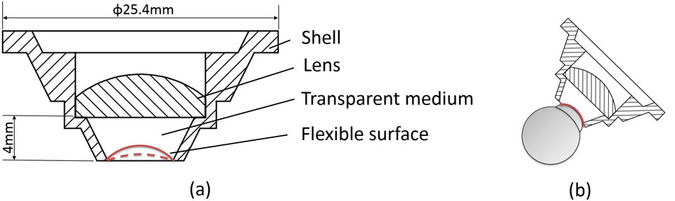
Fig. 1. Schematic design of the probe. (a) The structure of the contact probe for rats; (b) the surface of the soft gel can slightly change shape to adapt to different rodent eyes.
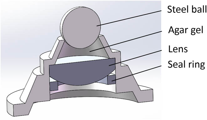
Fig. 2. Making the concave surface with a steel ball.
Fig. 3. Comparison between the flexible contact probe (a, c) and plano–concave method (b, d) in the spot diagram and PSF.
Fig. 4. Layout of the CSLO system for rodents, and a photo of the contact probe with the subject. L: lens, DM: dichroic mirror, PBS: polarizing beam splitter, QWP: quarter wave plate.
Fig. 5. In vivo retinal imaging. Comparison of the reflectance image by (a) the flexible contact probe and (b) the plano–concave lens for a rat; (c) autofluorescence imaging and (d) FA for the same rat; (e) reflectance and (d) fluorescence imaging for amouse. Scale bar: 100 μm.
|
Table 1. Ocular Parameters for Human, Rat, and Mouse Eyes[22]


Set citation alerts for the article
Please enter your email address

© Copyright 2018-2021 | Chinese Laser Press. All Rights Reserved 沪ICP备15018463号-20