• Acta Photonica Sinica
  • Vol. 50, Issue 8, 0850211 (2021)
Simin WU1, Bohan ZHANG1, Bin ZHENG3, and Minbiao JI1、2
Author Affiliations
  • 1State Key Laboratory of Surface Physics,Department of Physics,Fudan University,Shanghai200433, China
  • 2Yiwu Research Institute of Fudan University,Yiwu, Zhejiang3000, China
  • 3Zhejiang Provincial People's Hospital, Hangzhou10014, China
  • show less
    DOI: 10.3788/gzxb20215008.0850211 Cite this Article
    Simin WU, Bohan ZHANG, Bin ZHENG, Minbiao JI. Pump-probe Microscopy: Applications in Biomedicine and Materials Science (Invited)[J]. Acta Photonica Sinica, 2021, 50(8): 0850211 Copy Citation Text show less
    Three major processes in pump-probe process
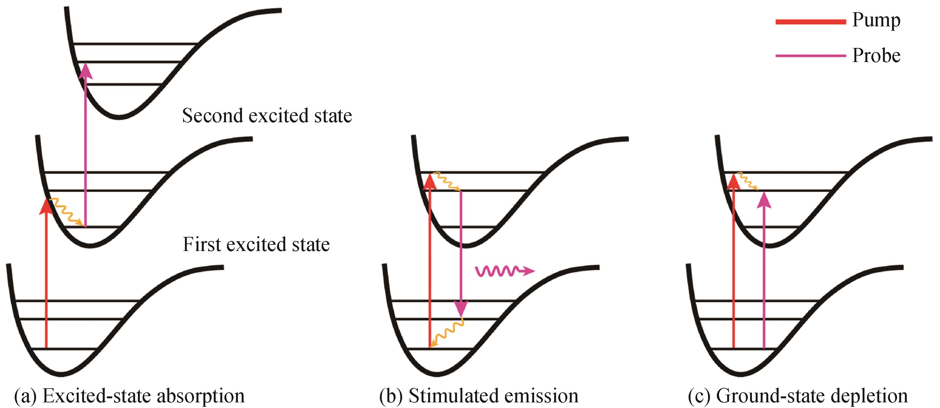
    Fig. 1. Three major processes in pump-probe process
    Schematic illustration of pump probe microscopy
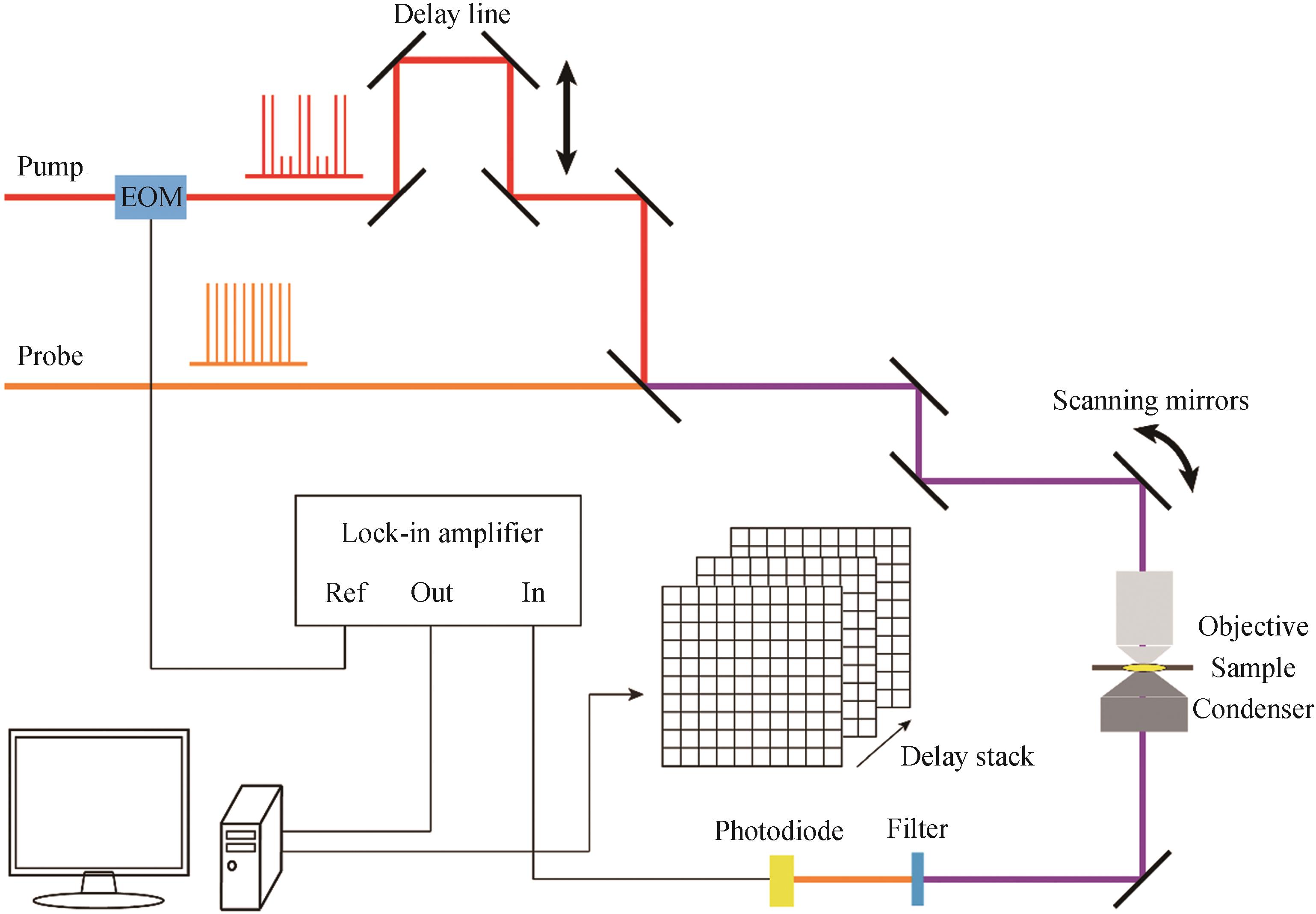
    Fig. 2. Schematic illustration of pump probe microscopy
    Imaging microvascular by pump-probe microscopy[46,56]
    Fig. 3. Imaging microvascular by pump-probe microscopy4656
    Characteristic transient optical responses of hemoglobin and hemosiderin[57]
    Fig. 4. Characteristic transient optical responses of hemoglobin and hemosiderin57
    Spectrally and temporally resolved TA spectra of hemoglobin and hemosiderin[57]
    Fig. 5. Spectrally and temporally resolved TA spectra of hemoglobin and hemosiderin57
    Melanoma tissue pump-probe signals[58]
    Fig. 6. Melanoma tissue pump-probe signals58
    Malignant melanoma compared with a benign nevus[58]
    Fig. 7. Malignant melanoma compared with a benign nevus58
    Pump-probe image of epitaxially grown graphene (Transient transmission data were probed at 780 nm following excitation at 390 nm. This data was recorded with a pump fluence of 14 mJ/cm2)[23]
    Fig. 8. Pump-probe image of epitaxially grown graphene (Transient transmission data were probed at 780 nm following excitation at 390 nm. This data was recorded with a pump fluence of 14 mJ/cm223
    Pump-probe results of monolayer MoS2[59]
    Fig. 9. Pump-probe results of monolayer MoS259
    Dual-modal SRS/TA imaging of MoS2 in live HeLa cells[63]
    Fig. 10. Dual-modal SRS/TA imaging of MoS2 in live HeLa cells63
    Few-layer BP samples and pump-probe microscopy[27]
    Fig. 11. Few-layer BP samples and pump-probe microscopy27
    Transient optical response of 4L-BP[27]
    Fig. 12. Transient optical response of 4L-BP27
    Transient dynamics of 8L-BP and layer-dependent resonances[27]
    Fig. 13. Transient dynamics of 8L-BP and layer-dependent resonances27
    Interlayer transition dynamics in WSe2/WS2 BL[83]
    Fig. 14. Interlayer transition dynamics in WSe2/WS2 BL83
    Interlayer coupling-dependent charge transfer from WS2 to graphene[84]
    Fig. 15. Interlayer coupling-dependent charge transfer from WS2 to graphene84
    Electron and energy transfer from tetracene (Tc) to WS2[85]
    Fig. 16. Electron and energy transfer from tetracene (Tc) to WS285
    Transient absorption image of M-SWNTs, S-SWNTs and DNA-SWNTs in cells[88]
    Fig. 17. Transient absorption image of M-SWNTs, S-SWNTs and DNA-SWNTs in cells88
    Time-resolved SSPP microscopy images[11]
    Fig. 18. Time-resolved SSPP microscopy images[11]
    Hot-carrier transport in hybrid perovskites within the first picosecond[35]
    Fig. 19. Hot-carrier transport in hybrid perovskites within the first picosecond35
    Simin WU, Bohan ZHANG, Bin ZHENG, Minbiao JI. Pump-probe Microscopy: Applications in Biomedicine and Materials Science (Invited)[J]. Acta Photonica Sinica, 2021, 50(8): 0850211
    Download Citation