
Wenbin Wang, Canbiao Li, Chujun Zheng. Retinal Blood Vessel Segmentation Using Hessian Based Orientational Adaptive Gabor Wavelet[J]. Laser & Optoelectronics Progress, 2020, 57(8): 081023
- Laser & Optoelectronics Progress
- Vol. 57, Issue 8, 081023 (2020)
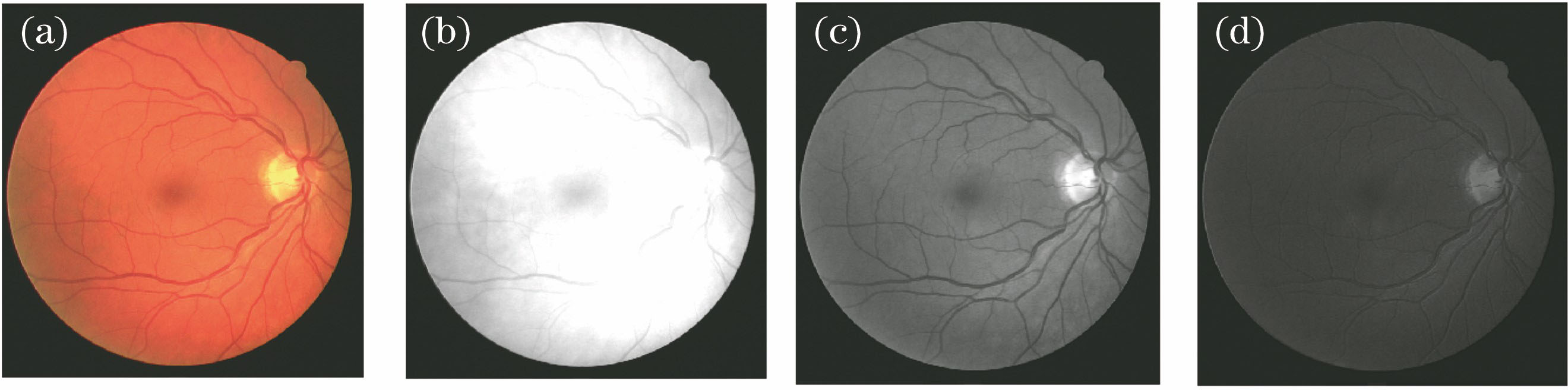
Fig. 1. Comparison of extracted results of green channel and other channels. (a) Color fundus image; (b) red channel; (c) green channel; (d) blue channel
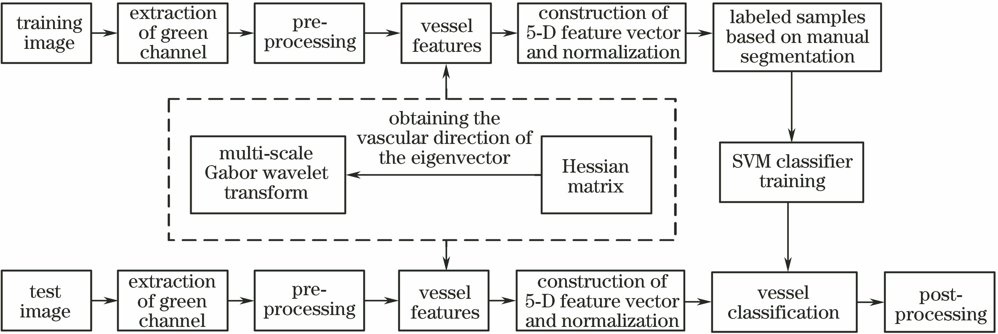
Fig. 2. Flow chart of proposed method
Fig. 3. Images of pre-processing results. (a) Color fundus image; (b) green channel; (c) expansion of region of interest in Fig. 3 (b); (d) image enhancement of Retinex in Fig. 3 (c)
Fig. 4. Normalized results of Hessian matrix feature. (a) Color fundus image; (b) fundus image of green channel of Fig. 4 (a); (c) maximal eigenvalue feature image of Hessian matrix of Fig. 4 (b)
Fig. 5. Normalized results of Gabor wavelet transform features at different scales. (a) a=2; (b) a=3; (c) a=4; (d) a=5
Fig. 6. Segmentation results of proposed method on DRIVE database. (a) Color fundus image; (b) fundus image of green channel of Fig. 6 (a); (c) expert manual segmentation image; (d) segmentation image of proposed method; (e) segmentation image of Gabor-SVM experiment
Fig. 7. Comparison of partial retinal segmentation images. (a) Complete segmentation image; (b)-(d) partial segmentation images of Fig. 7 (a)
|
Table 1. Performance comparison of blood vessel segmentation methods on DRIVE database

Set citation alerts for the article
Please enter your email address
© Copyright 2018-2021 | Chinese Laser Press. All Rights Reserved 沪ICP备15018463号-20