• Chinese Optics Letters
  • Vol. 20, Issue 10, 100003 (2022)
Xuanke Zeng1, Congying Wang1, Yi Cai1, Qinggang Lin1、2, Xiaowei Lu1、*, Jiahe Lin1, Xinming Yuan1, Wenhua Cao2, Yuexia Ai2, and Shixiang Xu1、**
Author Affiliations
  • 1Key Laboratory of Optoelectronic Devices and Systems of Ministry of Education and Guangdong Province, Shenzhen Key Laboratory of Micro-Nano Photonic Information Technology, College of Physics and Optoelectronic Engineering, , Shenzhen 518060, China
  • 2College of Electronics and Information Engineering, , Shenzhen 518060, China
  • show less
    DOI: 10.3788/COL202220.100003 Cite this Article Set citation alerts
    Xuanke Zeng, Congying Wang, Yi Cai, Qinggang Lin, Xiaowei Lu, Jiahe Lin, Xinming Yuan, Wenhua Cao, Yuexia Ai, Shixiang Xu. High spatial-resolution biological tissue imaging in the second near-infrared region via optical parametric amplification pumped by an ultrafast vortex pulse [Invited][J]. Chinese Optics Letters, 2022, 20(10): 100003 Copy Citation Text show less
    Schematic of the spiral PC idler imaging system.
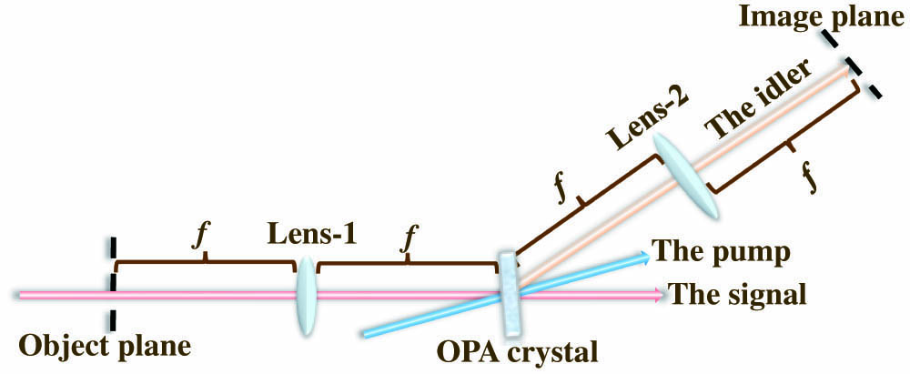
    Fig. 1. Schematic of the spiral PC idler imaging system.
    Theoretical simulations: (a) the spiral PC idler image of a phase-only object, (b) the traditional spiral PC image (without OPA), the idler images (c) with a Gaussian laser pump and (d) with a vortex laser pump of a quasi-phase object.
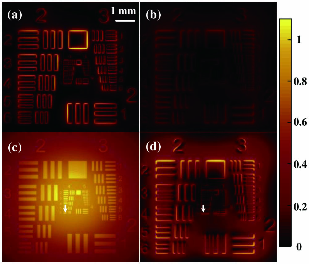
    Fig. 2. Theoretical simulations: (a) the spiral PC idler image of a phase-only object, (b) the traditional spiral PC image (without OPA), the idler images (c) with a Gaussian laser pump and (d) with a vortex laser pump of a quasi-phase object.
    Setup of the spiral PC idler imaging. VOA, variable optical attenuator; M, mirror; TA, target; MS, microscope; L1–L5, optical lenses; DM, dichroic mirror; TDL, time delay line; SPP, spiral phase plate; NC, OPA crystal; CCD, CCD camera.
    Fig. 3. Setup of the spiral PC idler imaging. VOA, variable optical attenuator; M, mirror; TA, target; MS, microscope; L1–L5, optical lenses; DM, dichroic mirror; TDL, time delay line; SPP, spiral phase plate; NC, OPA crystal; CCD, CCD camera.
    Idler images: (a1) and (b1) with Gaussian pump, (a2) and (b2) with vortex pump, and (c1) and (c2) for the local 1D intensity profile of Group 4.4 marked with the white arrow in (a1) and (a2), respectively. Meanwhile, (a1) and (a2) for an intensity object (a USAF 1951 resolution testing pattern) and (b1) and (b2) for a phase object (a pattern formed by a piece of flat glass coated with UV curing adhesive).
    Fig. 4. Idler images: (a1) and (b1) with Gaussian pump, (a2) and (b2) with vortex pump, and (c1) and (c2) for the local 1D intensity profile of Group 4.4 marked with the white arrow in (a1) and (a2), respectively. Meanwhile, (a1) and (a2) for an intensity object (a USAF 1951 resolution testing pattern) and (b1) and (b2) for a phase object (a pattern formed by a piece of flat glass coated with UV curing adhesive).
    Idler images of the USAF 1951 resolution testing pattern with a 40× MS: (a) Gaussian laser pump and (b) vortex laser pump.
    Fig. 5. Idler images of the USAF 1951 resolution testing pattern with a 40× MS: (a) Gaussian laser pump and (b) vortex laser pump.
    Microscopic images of frog egg cells and onion epidermis by (a1) and (b1) conventional MS imaging, (a2) and (b2) bright-field idler imaging, and (a3) and (b3) spiral PC idler imaging.
    Fig. 6. Microscopic images of frog egg cells and onion epidermis by (a1) and (b1) conventional MS imaging, (a2) and (b2) bright-field idler imaging, and (a3) and (b3) spiral PC idler imaging.
    Xuanke Zeng, Congying Wang, Yi Cai, Qinggang Lin, Xiaowei Lu, Jiahe Lin, Xinming Yuan, Wenhua Cao, Yuexia Ai, Shixiang Xu. High spatial-resolution biological tissue imaging in the second near-infrared region via optical parametric amplification pumped by an ultrafast vortex pulse [Invited][J]. Chinese Optics Letters, 2022, 20(10): 100003
    Download Citation