- Photonics Research
- Vol. 12, Issue 2, 271 (2024)
References
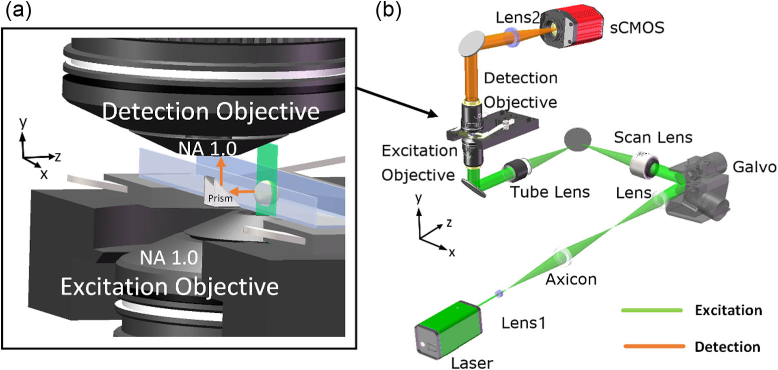

Yue Wang, Dashan Dong, Wenkai Yang, Renxi He, Ming Lei, Kebin Shi, "Reflective ultrathin light-sheet microscopy with isotropic 3D resolutions," Photonics Res. 12, 271 (2024)
Download Citation


Set citation alerts for the article
Please enter your email address

© Copyright 2018-2021 | Chinese Laser Press. All Rights Reserved 沪ICP备15018463号-20