
Zihan Wang, Li Zhang, Borui Zhou. Tomography system based on laser feedback confocal[J]. Infrared and Laser Engineering, 2020, 49(8): 20190541
- Infrared and Laser Engineering
- Vol. 49, Issue 8, 20190541 (2020)
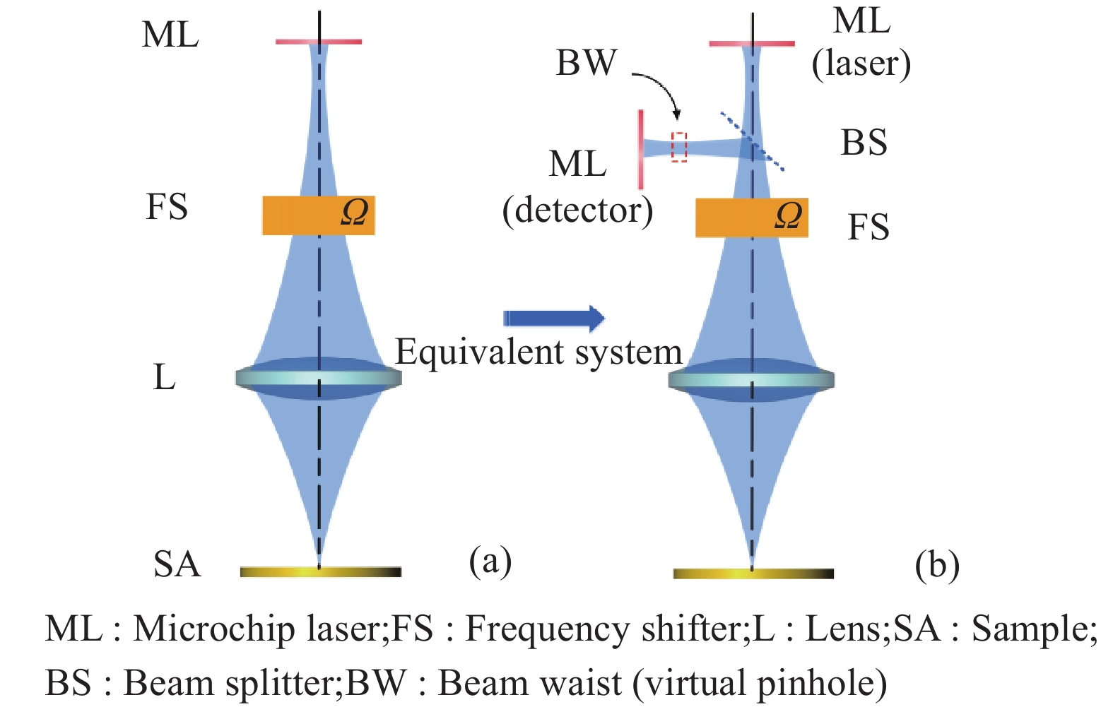
Fig. 1. (a) Principle diagram of LFCT and (b) equivalent confocal system
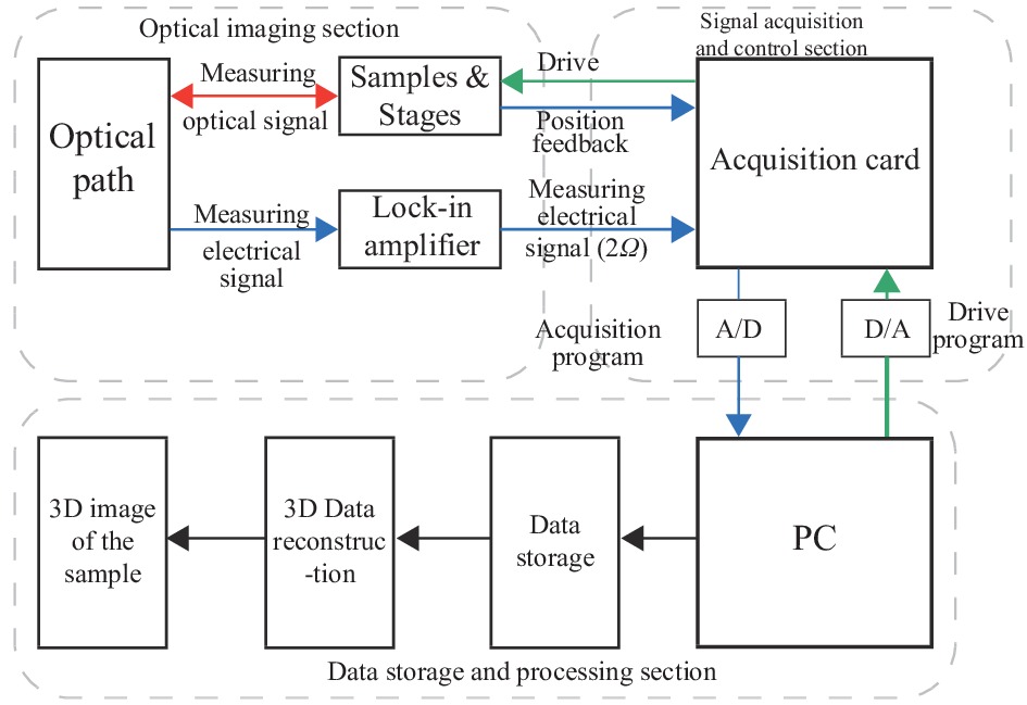
Fig. 2. Overall structure diagram of LFCT system
Fig. 3. Optical imaging section diagram of the LFCT system
Fig. 4. 3D scanning mode of LFCT system
Fig. 5. Flow diagram of positioning along the Z axis
Fig. 6. Single-dimensional data acquisition and step-driven LabVIEW program code
Fig. 7. Flow diagram of 3D isosurface display
Fig. 8. (a) Ruled grating measurement result of LFCT; (b) Defocus responding curve of LFCT
Fig. 9. [in Chinese]
Fig. 9. LFCT image and dark field microscope image of fresh onion innerepidermal tissue. (a) Image of dark field microscopic; (b) 3D image of LFCT; (c) Slice map of LFCT; (d) Contour map of LFCT
Fig. 10. Large-scale LFCT image of 500 μm depth inside fresh onion tissue


Set citation alerts for the article
Please enter your email address

© Copyright 2018-2021 | Chinese Laser Press. All Rights Reserved 沪ICP备15018463号-20