• Acta Photonica Sinica
  • Vol. 49, Issue 9, 0918001 (2020)
Sha AN1、2, Dan DAN1, Xiang-hua YU1, Tong PENG1、2, and Bao-li YAO1、2、*
Author Affiliations
  • 1State Key Laboratory of Transient Optics and Photonics, Xi'an Institute of Optics and Precision Mechanics, Chinese Academy of Sciences, Xi'an 710119, China
  • 2University of Chinese Academy of Sciences, Beijing 100049, China
  • show less
    DOI: 10.3788/gzxb20204909.0918001 Cite this Article
    Sha AN, Dan DAN, Xiang-hua YU, Tong PENG, Bao-li YAO. Progress and Prospect of Research on Single-molecule Localization Super-resolution Microscopy(Invited Review)[J]. Acta Photonica Sinica, 2020, 49(9): 0918001 Copy Citation Text show less
    Schematic diagram of various photoswitching modes of fluorescence proteins
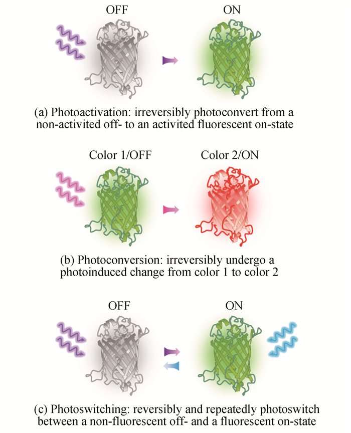
    Fig. 1. Schematic diagram of various photoswitching modes of fluorescence proteins
    Schematic diagram of single-molecule localization
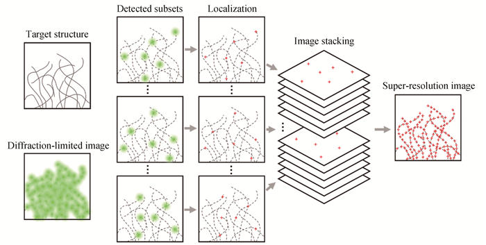
    Fig. 2. Schematic diagram of single-molecule localization
    Principle of PALM[26]
    Fig. 3. Principle of PALM[26]
    Principle of STORM with photo-switchable fluorophores[27]
    Fig. 4. Principle of STORM with photo-switchable fluorophores[27]
    Principle of 3D STORM[48]
    Fig. 5. Principle of 3D STORM[48]
    Schematic of Exchange-PAINT and super-resolution results[57]
    Fig. 6. Schematic of Exchange-PAINT and super-resolution results[57]
    Imaging implementation of 2D MINFLUX super-resolution nanoscopy[58]
    Fig. 7. Imaging implementation of 2D MINFLUX super-resolution nanoscopy[58]
    Different illumination modalities of fluorescence microscopy
    Fig. 8. Different illumination modalities of fluorescence microscopy
    Applications of SMLM in cytobiology
    Fig. 9. Applications of SMLM in cytobiology
    Comparison of axial plane diffraction-limited and corresponding super-resolution images of different targets[109]
    Fig. 10. Comparison of axial plane diffraction-limited and corresponding super-resolution images of different targets[109]
    Applications of SMLM in tissue biology
    Fig. 11. Applications of SMLM in tissue biology
    Applications of SMLM in neuroscience
    Fig. 12. Applications of SMLM in neuroscience
    SMLM methodResolutionFluorophoresDetected photon countsReferences
    PALM/FPALM~10~80 nmPhotoactivatable fluorescent proteinsLow (several hundred)[26], [28]
    STORM/dSTORM~20~50 nmPhotoswitchable organic dyesMedium (several thousand)[27], [30]
    QSTORM~24 nmQuantum dotsHigh (several thousand)[53]
    DNA-PAINT~5 nmAny single-molecule-compatible dyeExtremely high(hundreds of thousands)[54], [55], [56]
    MINFLUX~2 nmAny single-molecule-compatible dyeLow (zero toseveral hundred)[58], [59], [60]
    Table 1. Comparison of several SMLM methods
    Camera typeQuantumefficiencyReadoutnoisePixel numberPixel sizeFrame rateat full resolution
    Andor iXon3 897 EMCCD>90%< 1 e-512×51216 μm×16 μm35 fps
    Andor iXon Ultra 897 EMCCD>90%< 1 e-512×51216 μm×16 μm56 fps
    Hamamatsu Orca-Flash 4.0 LT sCMOS82%1.3 e-2 048×2 0486.5 μm×6.5 μm30 fps
    Hamamatsu Orca-Flash 4.0 V3 sCMOS82%1.0 e-2 048×2 0486.5 μm×6.5 μm100 fps
    Photometrics Prime 95B sCMOS95%1.6 e-1 200×1 20011 μm×11 μm41 fps
    Photometrics Prime BSI sCMOS95%1.6 e-2 048×2 0486.5 μm×6.5 μm43.5 fps
    Table 2. Comparison of main parameters between certain EMCCD and sCMOS
    Sha AN, Dan DAN, Xiang-hua YU, Tong PENG, Bao-li YAO. Progress and Prospect of Research on Single-molecule Localization Super-resolution Microscopy(Invited Review)[J]. Acta Photonica Sinica, 2020, 49(9): 0918001
    Download Citation