It is well-known that subcellular organelles are essential components of cells. Their morphological structures and dynamic characteristics directly reflect the physiological state of cells. Scientists have paid significant attention to the observation and analysis of the fine structures of subcellular organelles in living specimens.
The emerging super-resolution microscopy (SRM) techniques in the early 21st century, such as structured illumination microscopy (SIM), stimulated emission depletion (STED), and single-molecule localization microscopy (SMLM), skillfully bypass the limitation of the optical diffraction limit and effectively retain the advantages of optical microscopy. SRM techniques have been widely used in monitoring subcellular organelles in living cells.
This article systematically elaborates and analyzes the super-resolution structure characteristics of subcellular organelles in living cells. First, it briefly introduces the basic principles and fundamental characteristics of the three kinds of SRM techniques, i.e., STED, SIM, and SMLM, and expounds their development status. Second, the super-resolution fine structures and dynamic characteristics of subcellular organelles, such as the nucleus, cytoskeleton, mitochondrion, and endoplasmic reticulum (ER), are presented.
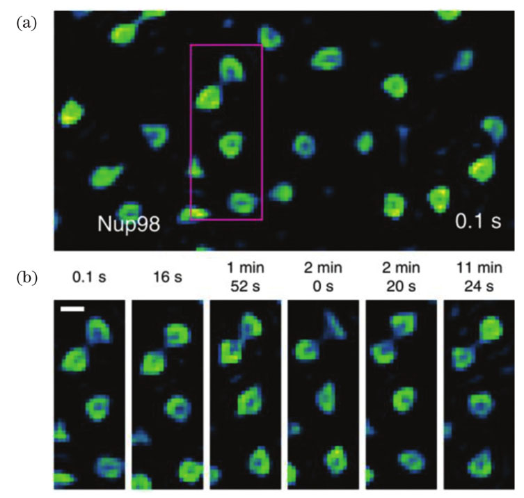
In 2016, Chagin et al. quantitatively measured and analyzed replication foci (RF) in mammalian cells using three-dimensional (3D) SIM. Mitchell-Jordan et al. (2012) directly imaged histone protein H3 in mammalian cells using STED to show the chromatin domain characteristics at the scale of 40-70 nm. Wombacher et al. and Lukinavicˇius et al. also employed STORM to observe the distribution of histone protein H2B in living HeLa and U2OS cells, respectively. Pelicci et al. (2020) imaged nuclear Lamin-A in intact nuclei of living cells through SPLIT-STED. Otsuka et al. (2016) captured images of different steps involved in assembling the NPC in a human cell. Lu et al. and Zhao et al. also realized the NPC super-resolution fluorescence imaging using different methods.
Gustafsson et al. (2009) employed SIM to monitor the dynamic characteristics and fine structures of microtubules. Additionally, Li Dong et al. further investigated the fine structures of the cytoskeleton based on SIM. Shao et al. (2011) clearly observed that microtubules in Drosophila S2 cells showed wrapped reticular structure and were distributed sparsely in these 3D-SIM images. Zhuang Xiaowei et al. (2012) revealed the 3D ultrastructure of the microfilament skeleton using the dual-objective STORM (Fig. 2). D’Este (2015) combined the two-color STED nanoscopy with SiR-Actin to show that the periodic cytoskeleton organization is ubiquity in axons and dendrites of living hippocampal neurons. Lukinavicˇius et al. (2014) disclosed the ninefold symmetry of the centrosome and the spatial organization of the actin in the axons of rats using STED. Recently, Wang et al. (2022) proposed JSFR-SIM and followed the microtubule motion in live COS-7 cells.
Additionally, Shim et al. (2012) observed the dynamic processes of mitochondrial fission/fusion through the STORM images of the mitochondrial membrane of living BS-C-1 cells. In 2020, the image data, hyperfine structures of mitochondria, and dynamic processes at different time points in living HeLa cells were attainable using STORM. Huang et al. (2018) successfully found the changes in mitochondrial crista during the fission and fusion of the mitochondrial. They identified the inter-cristae mergence in a single non-fusion mitochondrion using the Hessian-SIM system suitable for long-term super-resolution imaging of living cells (Fig. 3). Guo et al. (2018) also combined multicolor imaging technology with the newly proposed GI-SIM to observe the mitochondrial fission/fusion events at the ER-mitochondria contact sites. Wang et al. (2019) and Yang et al. (2020) accomplished the dynamic monitoring of mitochondrial crista using STED (Fig. 4). Recently, Wang et al. (2022) visualized the mitochondrial dynamics of living COS-7 cells through JSFR-SIM. A mitochondrion extended a tubulation tip, made contact with another mitochondrion, and then immediately retreated in the opposite direction.
Furthermore, Shim et al. (2012) successfully realized the STORM dynamic imaging of ER membrane (Fig. 5) and expressly observed the previously obscured details of morphological changes in ER remodeling. Georgiades et al. (2017) quantitatively analyzed the length and diameter of ER tubules using STORM. Guo et al. (2018) employed GI-SIM to obtain the formation and disappearance of ER contraction sites and the reconstruction of ER tubules in living COS-7 cells. Zhu et al. (2020) recently realized the real-time STED monitoring of 3D dynamic interaction between ER and mitochondria.
Finally, the development potential of combining super-resolution imaging with machine learning in exploring the fine structures of subcellular organelles is discussed.
It is an inevitable trend in cell image processing fields to apply deep learning algorithms in extracting information from subcellular super-resolution fluorescence images and help researchers analyze the image data. To achieve accurate and robust subcellular super-resolution image analysis, it is necessary to solve the problems of insufficient standardization of datasets and poor generalization ability of algorithm models.